The CEMC offers training for and access to the following instruments: Transmission Electron Microscope, Ultrathin Microtome, Vibratome, Olympus Confocal Microscope, Zeiss Confocal Microscope, and Image Analysis Workstation. The CEMC also has equipment marked with (*) for emerging technology development and currently only operated by CEMC staff.
Access to equipment
To schedule equipment use, please sign up through the E-Calendar on the NIDA/IRP SCIC homepage (link: https://bis.nida.nih.gov/home/ Requires NIDA IRP Network Access). For rescheduling or cancellations, please email shiliang.zhang@nih.gov.
FEI Tecnai G2 12 TEM with a Gatan OneView 16 Megapixel Camera
Location – BRC B1A323A
FEI Tecnai G2 12 Transmission Electron Microscope (TEM) with a Gatan OneView 16 Megapixel Camera is a TEM with an operating voltage range of 20 to 120 KV. Electron micrographs can be collected with this microscope with magnification of thin samples (<200 nm) up to 700,000 times. A Gatan OneView 16 Megapixel Camera is for digital image acquisition with imaging and in-situ data capture at full 4k × 4k resolution.
-
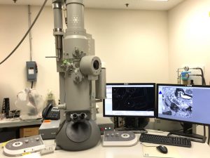
- FEI Tecnai G2 12 TEM with a Gatan OneView 16 Megapixel Camera

-

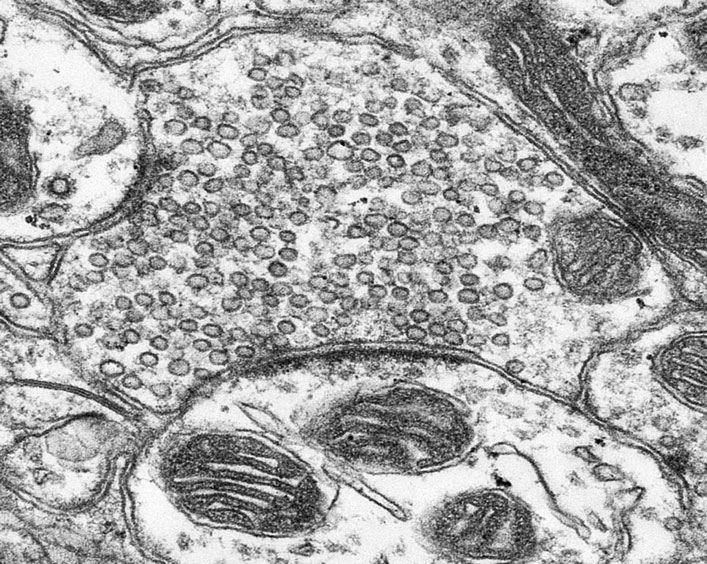
- An axon terminal containing synaptic vesicles and making synapses (TEM image taken with the FEI Tecnai TEM Microscope)
Zeiss LSM 980
Location – BRC B1A322
Zeiss LSM 980 with Airyscan 2 Confocal Microscope is optimized for simultaneous spectral detection of multiple weak labels with the highest light efficiency. The unique combination of spectral imaging with improved signal-to-noise ratio and resolution enables lower laser power for the live cell experiments. Airyscan 2 allows you to do more than any conventional LSM detector. Each of its 32 detector elements collects additional information, while all of them together gather even more light, yielding super-resolution quantitative results. By adding structural information with Joint Deconvolution (jDCV), you can push resolution even further. Or use the Multiplex modes to get super-resolution information up to 10 times faster. ZEN microscopy software puts a wealth of helpers at your command to achieve reproducible results in the shortest possible time. AI Sample Finder helps you quickly find regions of interest, leaving more time for experiments. Smart Setup supports you in applying best imaging settings for your fluorescent labels. Direct Processing enables parallel acquisition and data processing.
LSM 980 Upright Confocal Microscope is designed for high-quality of fixed samples. It features an impressive array of capabilities, including 8 laser lines (405, 445, 488, 514, 561, 594, 639 and 730 nm) that provide exceptional flexibility for various fluorophores and ensure optimal separation. With a 34-channel spectral array and 2 Near IR optimized detectors, spanning a wide imaging range from 390 to 900 nm, the microscope facilitates easy and precise separation of signals. It supports simultaneous collection of up to 12 channels, enhancing efficiency and throughput in imaging experiments. Moreover, the LSM 980 incorporates a super-resolution detector, the Airyscan, capable of achieving resolutions as fine as 90 nm. This advanced feature enables detailed and accurate visualization of biological structures at the molecular level.
-
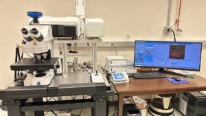
- Zeiss LSM 980

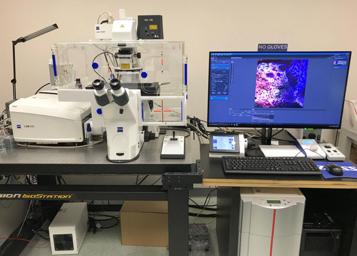
Zeiss LSM880 Airyscan/CY7.5 Confocal Microscope
Location – BRC B1A322
Zeiss LSM880 Airyscan/CY7.5 Confocal Microscope offers high sensitivity, enhanced resolution in x,y and z, and high image-acquisition speed in one system. Users achieve a 1.7× higher resolution in all spatial dimensions, 140 nm laterally and 400 nm axially. The improved sensitivity leads to better image quality and increased speed. The whole imaging process is possible with standard sample preparation and labeling protocols. With Airyscan, ZEISS introduces a new concept. Instead of detecting signals with a single point detector, a multichannel area detector with 32 elements collects all the light from an Airy disk simultaneously. Hereby, each detector element functions as a single, very small pinhole. Knowing the beam path and the spatial distribution of each Airy disk enables very light-efficient imaging: researchers can now use all photons that the objective collected.
-
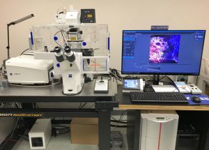
- Zeiss LSM880 Airyscan/CY7.5 Confocal Microscope
-
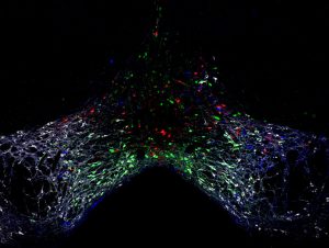
- Dopaminergic, glutamatergic and GABAergic neurons in the ventral tegmental area(Confocal image taken with the Zeiss LSM880 Airyscan/CY7.5 Confcoal Microscope)
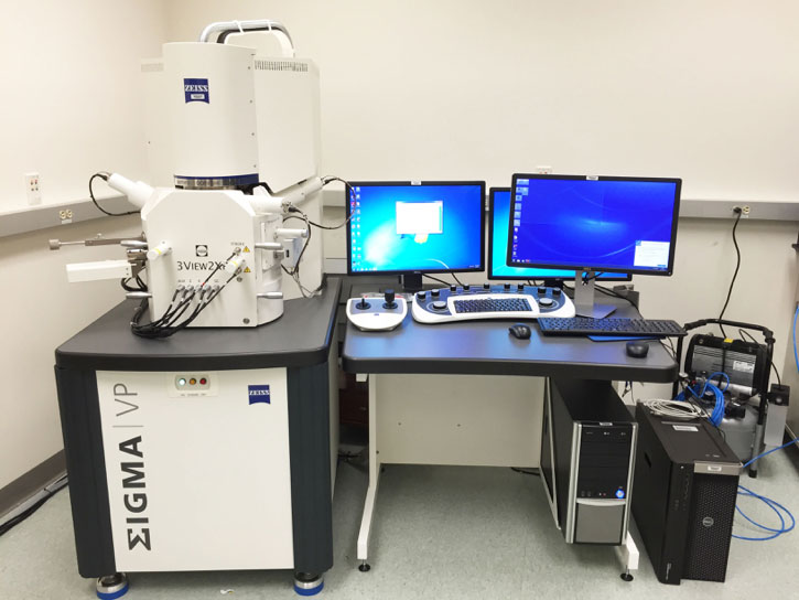
Zeiss Sigma VP Field Emission Scanning Electron Microscope (SEM) with Gatan 3View2 XP System
Location – BRC B1A323
* Zeiss Sigma VP Field Emission Scanning Electron Microscope (SEM) with Gatan 3View2 XP System is used for image collection for 3-D reconstruction of neurons. The 3-D SEM system has the follow properties: (a) ultrathin microtome inside the scanning electron microscope chamber allows repeated cutting and imaging; (b) cut thickness can be set at 30 nm, even less than 15 nm; (c) the maximum sample size is about 1 mm × 1 mm × 0.6 mm; (d) the efficiency is very high. A 100-slice dataset can be collected in a few hours. A 1000-slice dataset can be collected with an overnight run.
-

- Zeiss Sigma VP Field Emission Scanning Electron Microscope (SEM) with Gatan 3View2 XP System
-
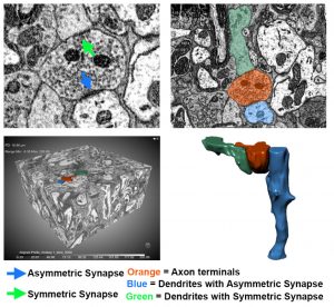
- 3-D reconstruction of a single axon terminal in the lateral habenula establishing asymmetric and symmetric synapses (SEM 3D image visualized with the DragonFly software)

Leica VT1000S Vibratome
Location – BRC 08A314.02
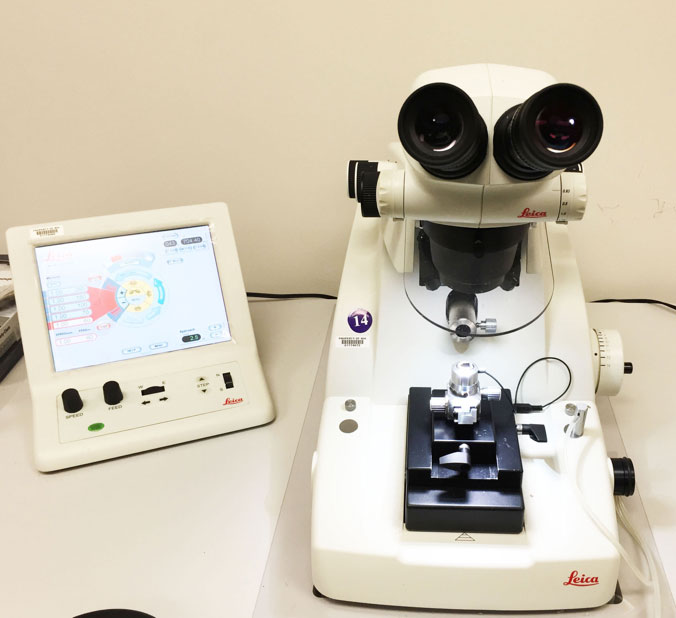
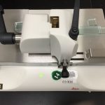
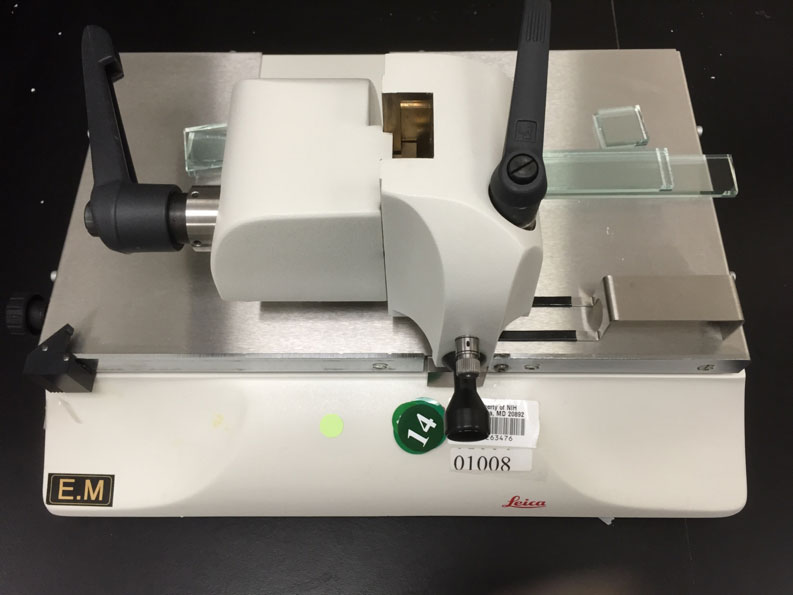
Leica UC7 Ultrathin Microtome
Location – BRC B1A326
Leica Glass Knife Maker
Location – BRC B1A326
Leica VT1000S Vibratome is used to section the whole brain in 30-200 µm thickness. Leica UC-7 Ultrathin Microtome is used to section samples in resin block in 60-70 nm thickness. Leica Glass Knife Maker is used to make glass knives for ultrathin sectioning before changing to a diamond knife.
-
- Leica VT1000S Vibratome

-
- Leica UC7 Ultrathin Microtome
-
- Leica Glass Knife Maker
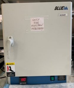
Blue M Regular Oven
Location – BRC 08A314.05
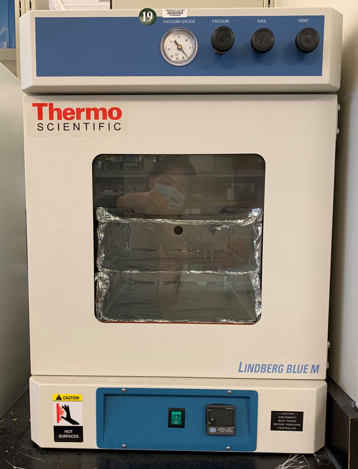
Lindberg Blue M Vacuum Oven
Location – BRC 08A314.05
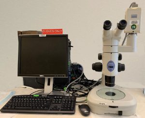
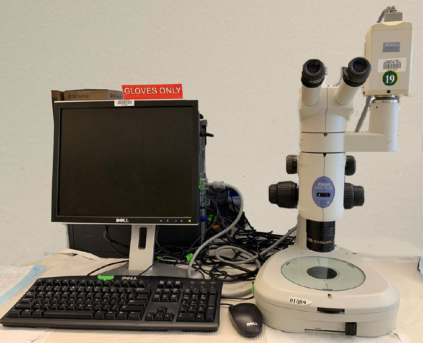
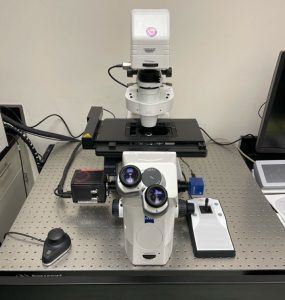
Nikon SMZ150 Dissecting Microscope
Location – BRC 08A314.05
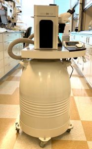
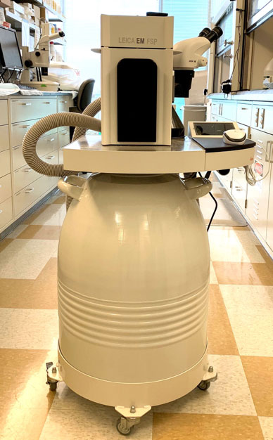
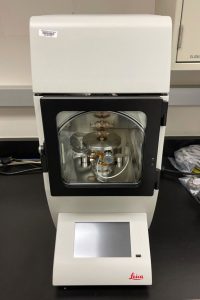
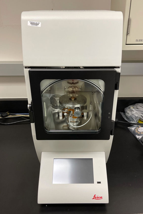
Leica EM AFS2
Location – BRC 08A314.05
Equipment for Tissue Processing: Blue M Regular Oven is used for pre-embedding resin polymerization. Lindberg Blue M Vacuum Oven is used for post-embedding resin polymerization. Nikon SMZ150 Dissecting Microscope is used to select the ROI of samples. Leica EM AFS2 performs freeze substitution and progressive lowering of temperature techniques and allows low temperature embedding and polymerization of resins.
-
- Blue M Regular Oven

-

- Lindberg Blue M Vacuum Oven
-
- Nikon SMZ150 Dissecting Microscope
-
- Leica EM AFS2
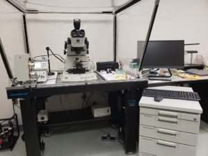
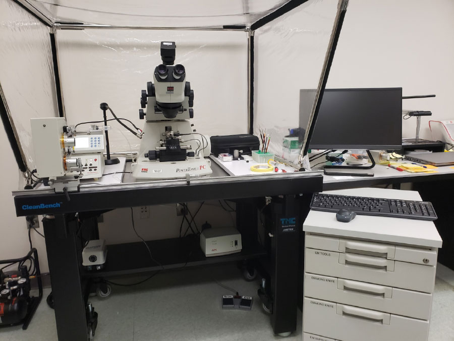
RMC Automatic Tape Collecting Ultramicrotome (ATUMtome) – B1A326
Location – B1A326
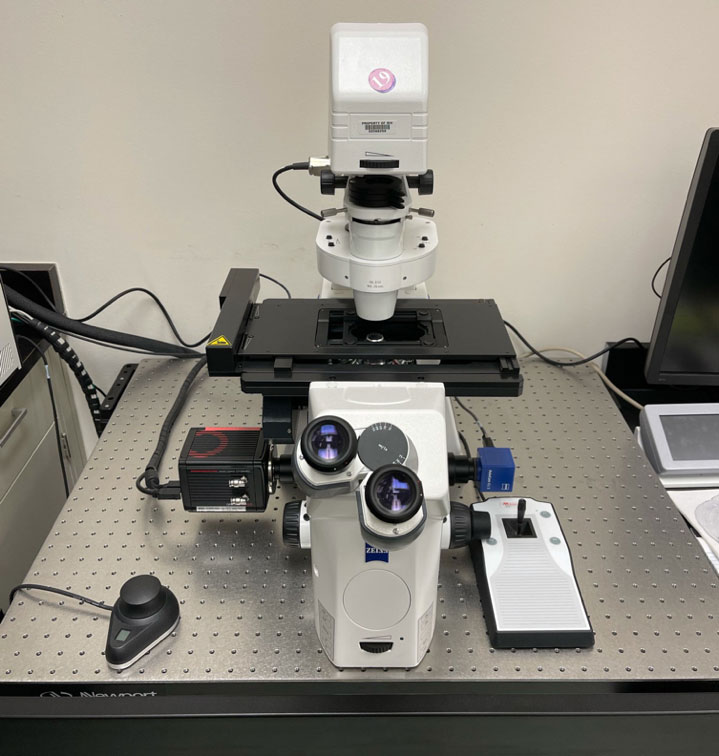
Zeiss AxioObserver Microscope
Location – BRC B1A326
Leica ACE600 Sputtering and Carbon Coater
Location – BRC B1A326
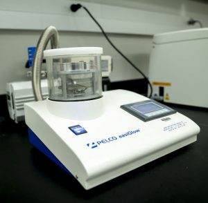
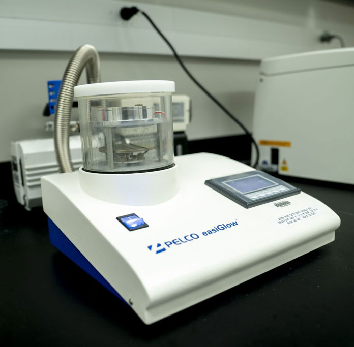
PELCO easiGlow™ Glow Discharge Cleaning System
Location – BRC B1A326
* Equipment for emerging technology development in Correlative Light and Electron Microscopy (CELM) – ATUMtome is used to collect hundreds to thousands of serial ultrathin sections onto to a tape substrate for later analysis and SEM imaging. Zeiss AxioObserver Microscope with ZEN Blue software is used to collect fluorescent images. Leica ACE600 Sputtering and Carbon Coater is used to produce thin, but strong support layers for SEM high-resolution imaging of structured non-conductive specimens. PELCO easiGlow™ Glow Discharge Cleaning System is primarily designed for cleaning TEM grids and preventing wrinkles in the polymer tape in CLEM.
-
- RMC Automatic Tape Collecting Ultramicrotome (ATUMtome)
-
- Zeiss AxioObserver Microscope
-
- Leica ACE600 Sputtering and Carbon Coater
-
- PELCO easiGlow™ Glow Discharge Cleaning System
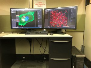
Data Analysis Workstations
Workstation A
Location – BRC08A205.07
Workstation A includes software: Imaris and Amira.
Workstation B
Location – BRC 08A205.08
Workstation B includes software: Huygens Deconvolution.
Image Analysis Workstation – Available software: Imaris, Amira, Dragonfly, Image J, Image Pro, Huygens Professional for deconvolution, Zen Lite, Adobe, etc.
-
- Data Analysis Workstation

