Access to equipment
Please request training on how to operate the equipment before scheduling. To schedule equipment use, sign up through the E-Calendar on the NIDA/IRP SCIC homepage (link: https://bis.nida.nih.gov/home/ Requires NIDA IRP Network Access). For rescheduling or cancellations, please email shiliang.zhang@nih.gov.
Olympus VS200 Scanner
Location: 08A314A

Olympus VS200 Scanner (location: 08A314A)
Features:
- Provides reliable images for quantitative data analysis and improves workflow efficiency.
- Scans standard histology slides and fluorescently labeled slides.
- Supports brightfield, fluorescence, darkfield, phase contrast, and polarization imaging.
- Objectives: 2x, 4x, 10x, 20x, 40x, 60x oil lens with accurate autofocus.
- Scans up to 210 slides in a batch with robotic loader.
- Automatic oil dispenser for high magnification.
- Flexible batch scan mode allows different observation methods per slide.
Miltenyi Biotec UltraMicroscope Blaze
Location: 08A314A
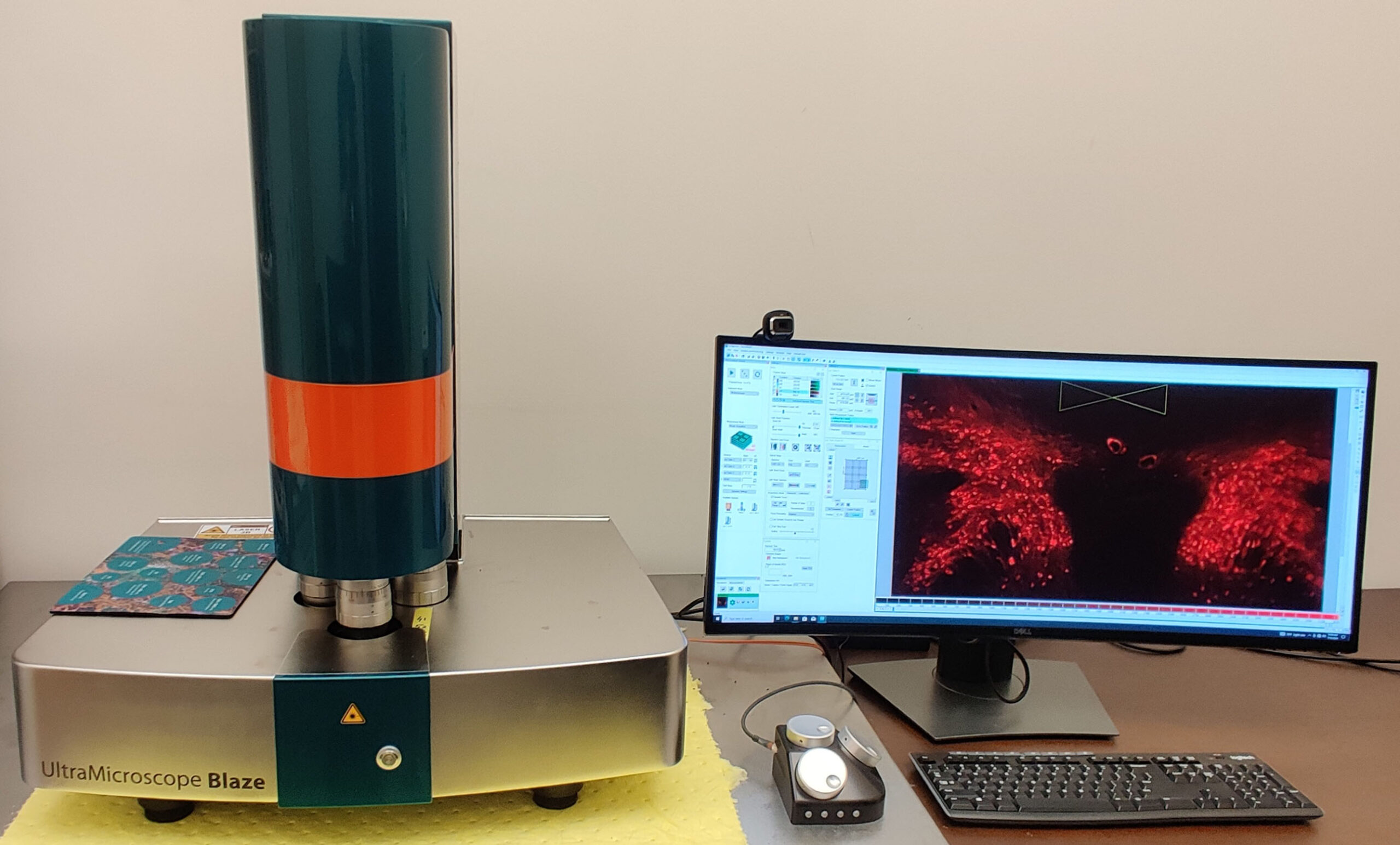
Miltenyi Biotec UltraMicroscope Blaze (location: B1A408)
Features:
- Advanced light sheet optics ensure excellent data quality.
- Fully automated system for imaging large or multiple cleared samples at subcellular resolution.
- Standard chamber for multiple rodent organs or organoids.
- XXL chamber accommodates very large samples including whole adult mouse/rat models.
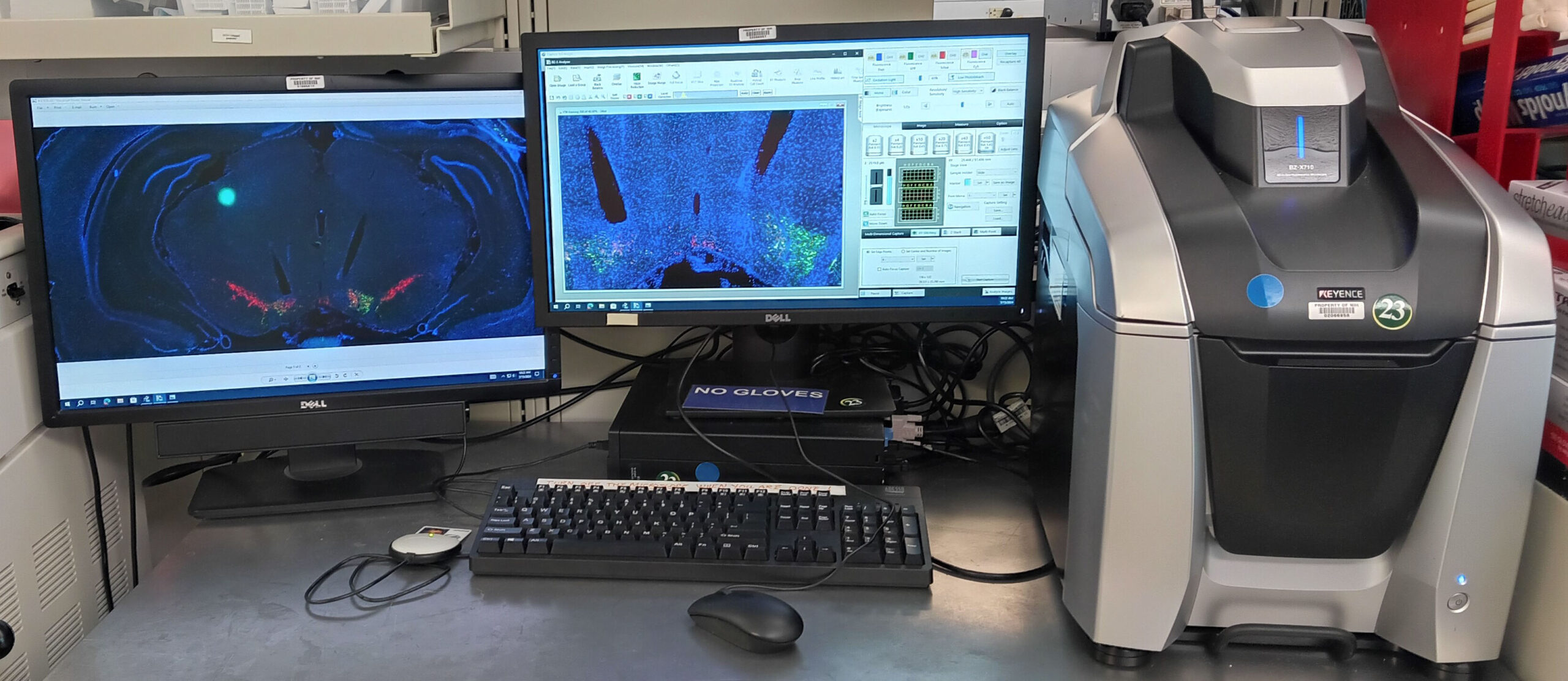
Keyence BZ-X710 Microscope
Location: 08A314.06
- Collects mosaic images using 2x, 4x, 10x, 20x, 40x, 60x objectives.
- Supports up to 4 fluorophores simultaneously.
- Images up to 3 slides at a time with excellent stitching and autofocus.
Olympus MVX710 Microscope
Location: 08A314A
- Dissection-style stereoscopic microscope ideal for injection site documentation.
- Objectives: 63x and 2x with 0.63x–6.3x secondary adjustments.
- Supports detection.
Keyence BZ-X720 Microscope
Location: 08A314.06
Keyence BZ-X720 Microscope (location: 05A314.10)
Features:
- Collects mosaic images using 2x, 4x, 10x, 20x, 40x, 60x objectives.
- Supports up to 4 fluorophores simultaneously.
- Images up to 3 slides at a time with excellent stitching and autofocus.
Olympus MVX10 Microscope
Location: 08A314A
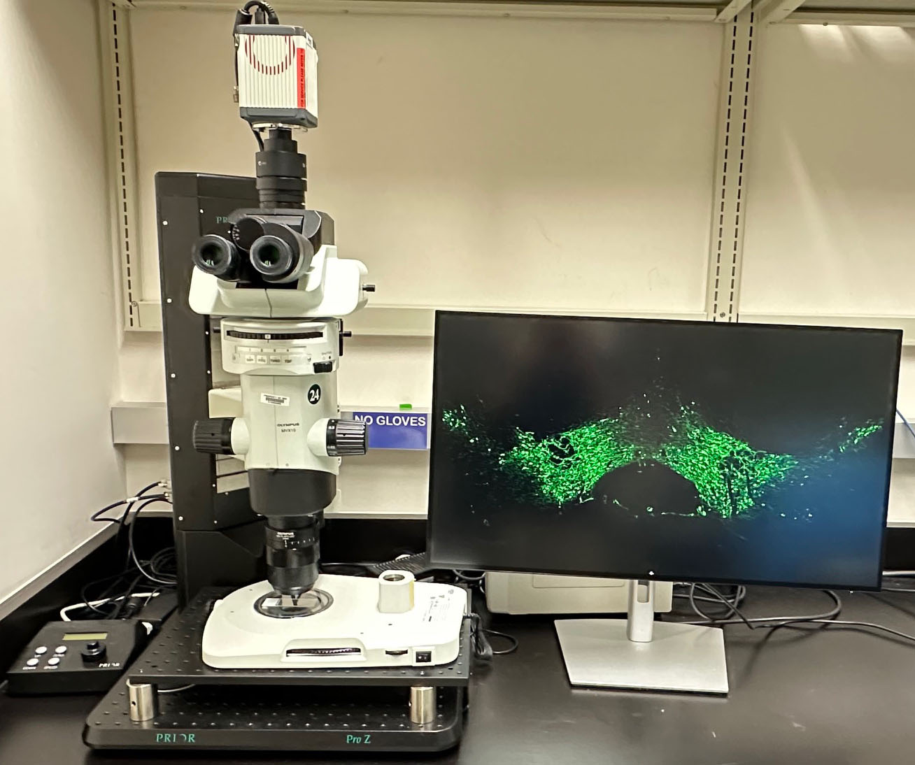
Olympus MVX10 Microscope (location: 08A314A)
Features:
- Dissection-style stereoscopic microscope ideal for injection site documentation.
- Objectives: 63x and 2x with 0.63x–6.3x secondary adjustments.
- Supports detection of up to 3 fluorophores including DAPI, GFP, FITC, tdTomato, CY5, and others.
Leica 3050S Cryostats
Leica 3050S Cryostat-1 – Location: 08A314.02
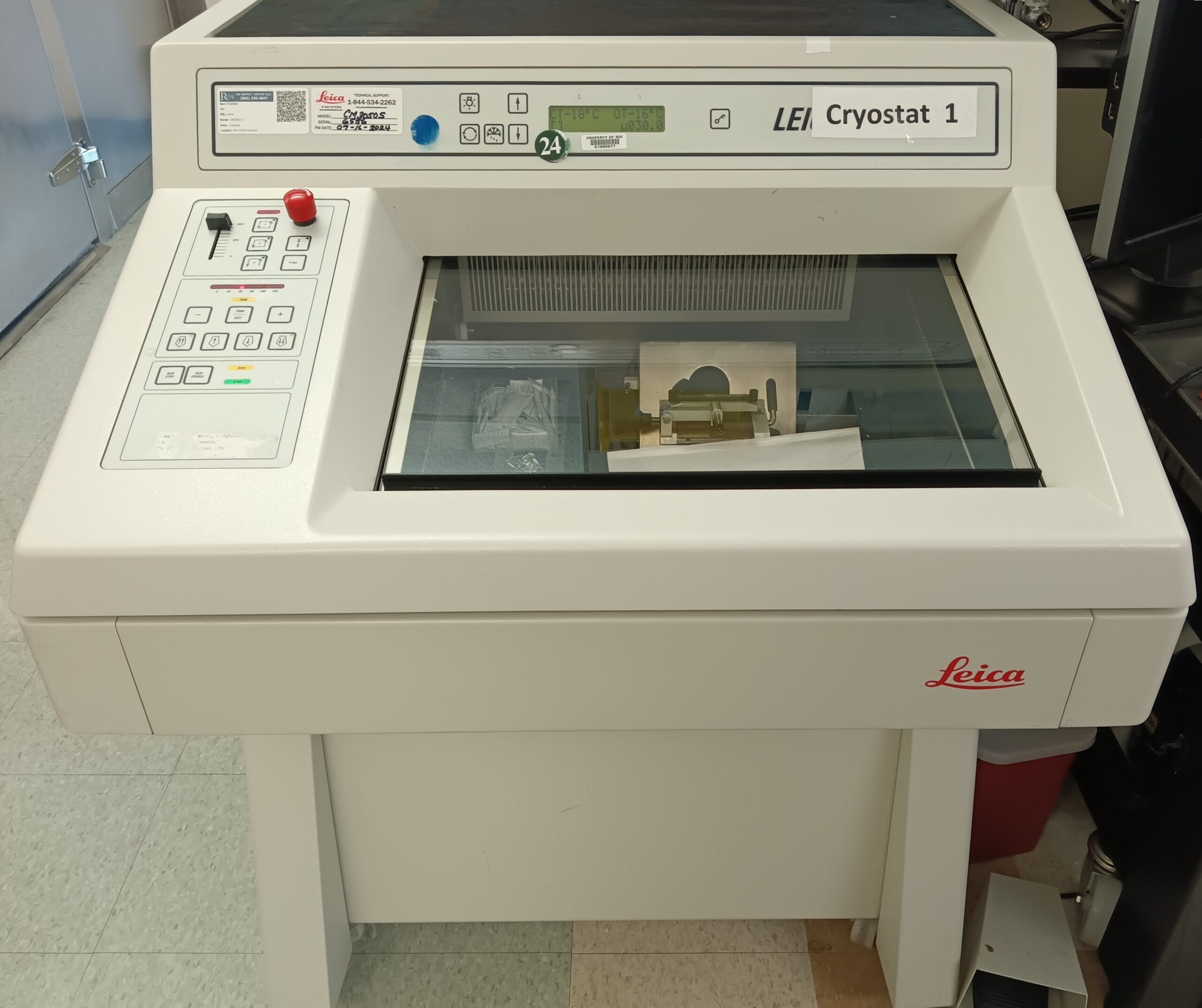
Leica 3050S Cryostat-1 (location: 05A314.10)
Leica 3050S Cryostat-2 – Location: 08A314.04
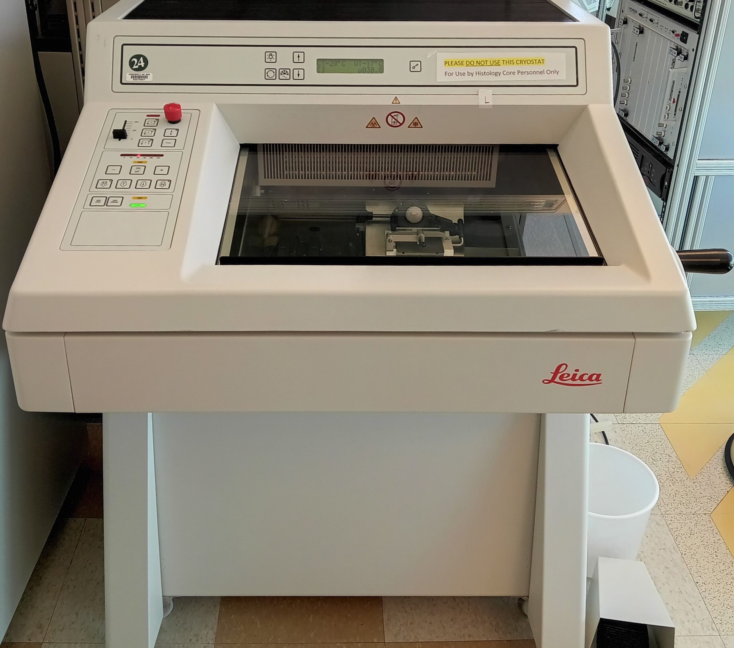
Leica 3050S Cryostat-2 (location: 05A314.10)
Features:
- Designed for sectioning samples at low temperature.
- Produces sections ranging from 5–50 μm thickness.
Leica Vibratome VS1000T
Location: 08A314.06
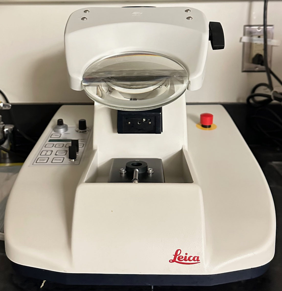
Leica Vibratome VS1000T (location: 05A314.7)
Features:
- Designed for sectioning fixed tissue specimens at room temperature.
- Produces sections ranging from 30–250 μm thickness.
- Consistently generates thin sections, including difficult non-homogeneous specimens.
Data Analysis Workstations
-

- Computer A – Imaris (location: 08A205.10)
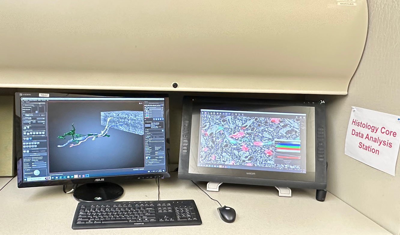
-
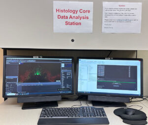
- Computer B – VS Export (location: 08A205.07)

Features:
- The Histology and Imaging Core provides two computers for open use by NIDA IRP researchers.
- Computer 1 – VS Export (Location: 08A205.09).
- Computer 2 – Imaris, Dragonfly (Location: 08A205.10).
