A NIDA IRP Core Facility
Acting Core Manager
Contact
Location:
Biomedical Research Center
251 Bayview Blvd
Suite 200, Room 08A709
Baltimore, MD 21224
Phone:443-740-2698
Email: shiliang.zhang@nih.gov
About the Core
The NIDA-IRP Histology and Imaging Core (HIC) serves NIDA-IRP researchers by providing access to existing and emerging neuroanatomical technologies to map and characterize neuronal circuits.
Core News:
- BIS has created an online application (Network or VPN required) for submitting quotes for contract services.
- Visual Media Services and the Histology and Imaging Core have teamed up to create training videos. Check out the Equipment page to see videos for the Keyence and Leica Thunder microscopes.
Goals and Services:
- Increase access to existing and emerging neuroanatomical techniques to NIDA-IRP researchers. We achieve this goal using both emergent technologies and classic methodologies. Our standard procedures include cryosectioning, multiplex immunohistochemistry and in situ mRNA hybridization, counterstaining (DAPI, cresyl violet, etc), widefield fluorescent and brightfield microscopy.
- Develop and adopt emerging neuroanatomical techniques that NIDA-IRP researchers can use. We are refining in situ hybridization for multiplex mRNA expression methods, large-volume tissue clearing and immunolabeling methodologies and advanced imaging techniques such as light-sheet microscopy. We are open to developing projects with NIDA-IRP investigators using these methods.
- Provide consultations and training to NIDA-IRP researchers in the adaptation and execution of neuroanatomical techniques and procedures.
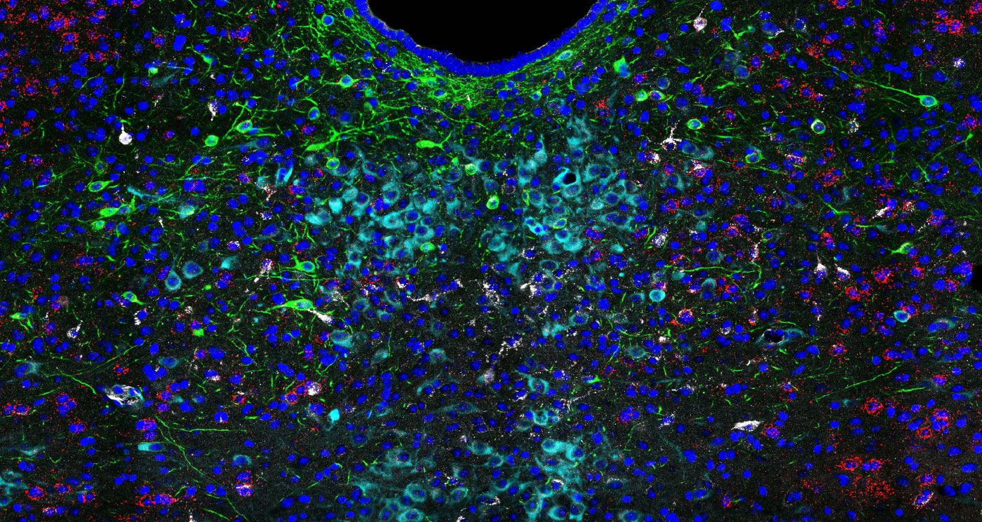
 We act as a resource with a diverse array of training tools including workshops, seminars, and one-on-one sessions. We are also able to consult investigators on image analysis and offer advanced analysis software for image rendering and quantification.
Please send an email to discuss usage of the core facilities or to start a new project!