High Spatiotemporal Resolution Radial Encoding Single-Vessel fMRI
Published in Advanced Science (Weinh).
Authors
Yuanyuan Jiang, Patricia Pais-Roldán, Rolf Pohmann and Xin Yu
Paper presented by Dr. Zilu Ma and selected by the NIDA TDI Paper of the Month Committee
Publication Brief Description
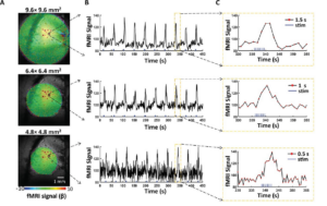
Conventional functional magnetic resonance imaging (fMRI) is developed to measure hemodynamic responses as a surrogate of neuronal activity. Recent development in single-vessel fMRI enables distinction of arteriole-dominated cerebral blood volume (CBV) and venule-dominated blood oxygenation level dependent (BOLD) fMRI signals from intracortical vessel voxels. However, increased spatial and temporal resolution of single-vessel fMRI acquisition leads to inevitable single-to-noise ratio (SNR) loss. Using methods such as reshuffled k-t space fast slow angle shot (FLASH) or balanced steady state free recession (fSSFP) sequences can ensure sufficient SNR, however, there remains a challenge to push resolution higher given the interdependent spatial, temporal resolution and SNR. This paper presents a radial encoding MRI scheme that can achieve the finest spatial scale for single-vessel fMRI to measure the individual vessels penetrating the rat somatosensory cortex. Radial encoding offers continuous updating of the center of the k-space and pushes the 50 × 50 μm2 spatial resolution with a 1 to 2 Hz sampling rate by defining the arbitrary number of projections in the azimuthal direction. Besides detecting refined hemodynamic maps of intracortical vessels, the radial encoding based single-vessel fMRI offers the opportunity to distinguish the intravascular and extravascular effects from the cortical vessels. This further benefits real-time single-vessel BOLD fMRI, CBV, and cerebral blood flow studies, making it a valuable tool for advanced brain functional mapping with high-field MRI scanners.
High Spatiotemporal Resolution Radial Encoding Single-Vessel fMRI Journal Article
In: Adv Sci (Weinh), vol. 11, no. 26, pp. e2309218, 2024, ISSN: 2198-3844.
