• Skip to primary navigation
  • Skip to main content
  • Skip to primary sidebar

NIDA IRP

National Institute on Drug Abuse - Intramural Research Program

  National Institute on Drug Abuse | NIH IRP | Treatment Info | Emergency Contacts
  • Home
  • News
    • Featured Paper of the Month
    • Reviews to Read
    • Hot off the Press
    • IRP News
    • Awards
    • Technology Development Initiative Paper of the Month
    • Seminar Series
    • Addiction Grand Rounds
  • About
    • About NIDA IRP
    • Contact Us
    • Directions and Map
    • Careers at NIDA IRP
    • Emergency Contacts
    • Employee Assistance Resources
  • Organization
    • Faculty
    • Office of the Scientific Director
    • Office of the Clinical Director
    • Office of Education and Career Development
    • Administrative Management Branch
    • Molecular Targets and Medications Discovery Branch
    • Cellular and Neurocomputational Systems Branch
    • Molecular Neuropsychiatry Research Branch
    • Neuroimaging Research Branch
    • Behavioral Neuroscience Research Branch
    • Integrative Neuroscience Research Branch
    • Translational Addiction Medicine Branch
    • Core Facilities
    • Community Outreach Group
  • Training Programs
    • Office of Education and Career Development
    • OECD Awards
    • Summer Internship Program
    • Postbaccalaureate Program
    • Graduate Partnership Program
    • Postdoctoral Program
    • NIDA Speakers Bureau
    • Clinical Electives Program
    • Clinical Mentoring Program
  • Study Volunteers
  • Transgenic Rat Home
  • OTTC Information
  • Publications
  • Technology Development Initiative
  • Links

LE-Tg(Gad1-iCre)3Ottc

Last Updated on November 12, 2024

Background | Status & Availability | Transgene Info | Phenotypic Characterization | Breeding | Genotyping | References | Blog/Comments/Reviews | Related rats | Acknowledgements

Background

Glutamate decarboxylase 1 (brain, 67kDa) (GAD67), also known as GAD1, is the enzyme responsible for catalyzing the conversion of L-glutamic acid into gamma-aminobutyric acid (GABA).  GABA is the primary inhibitory neurotransmitter in the mammalian brain and GABAergic neurons can be identified by expression of GAD1.  Not all GABAergic neurons express GAD1 (some use only GAD2).  We have generated and characterized a strain of transgenic Long Evans rats expressing iCre recombinase under a GAD1 promoter (GAD1::iCre, line 3).  The tissue-specific expression of iCre can be used in combination with Cre-dependent transgenes to obtain selective expression of transgenes in GAD1(+) neurons.

Status and Availability

This rat has been published (PMID: 28690111).

As of May 17, 2016, this strain is available as line 751 at the RRRC. You are now exiting the NIDA IRP Website

This rat is registered at the Rat Genome Database (RGD) as RGD ID#9588593. You are now exiting the NIDA IRP Website

Transgene Information

Figure 1:  A schematic of the Gad1-iCre transgene.
Figure 1:  A schematic of the Gad1-iCre transgene.  A bacterial artificial chromosome (BAC) containing the rat Gad1 gene (CH230-24D16) was obtained from CHORI, and recombineered to replace the start codon of Gad1 with a 2.6 kb cassette containing iCre (improved Cre recombinase), the polyadenylation signal from the gene for bovine growth hormone, and a galk bacterial selection marker (pOTTC335).  This BAC was injected into the pronuclei of fertilized Long Evans rat embryos by NIMH Transgenic Core, and ultimately resulted in 3 independent, phenotypically positive Gad1::iCre lines.  This line (LE-Tg(Gad1-iCre)3Ottc) has a single copy of the transgene per haploid genome as determined by droplet digital PCR.


Phenotypic Characterization

Figure 2: Basic Characterization of the GAD-Cre Rat.

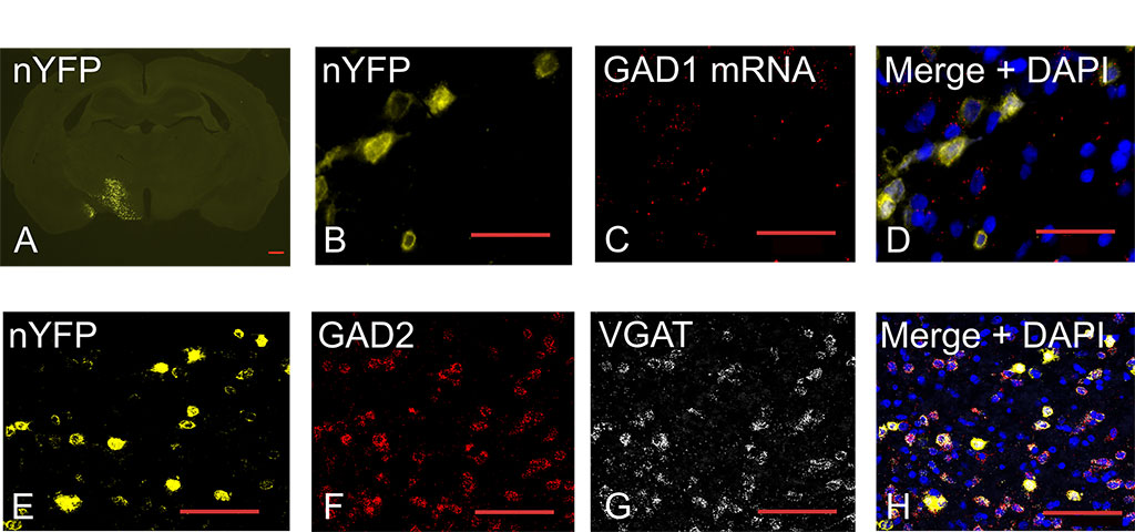
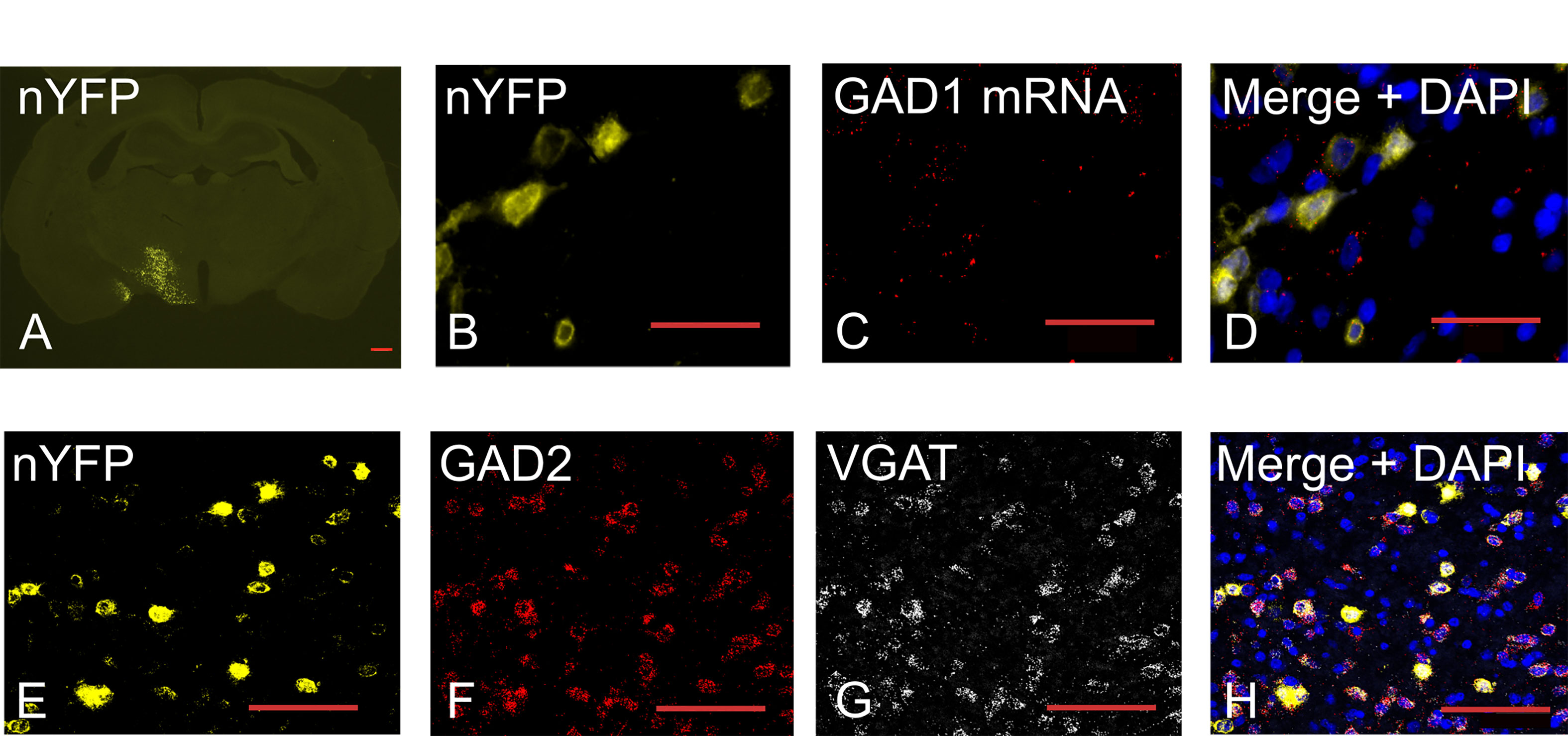
Figure 2:  Basic Characterization of the GAD-Cre Rat.  (A) Coronal section showing nuclear YFP (nYFP) expression in LH 2 weeks after AAV injection. The scale bar represents 0.5 mm.  Colocalization of (B) nYFP protein fluorescence (C) and GAD1 mRNA.  (D) Merged image of nYFP (yellow), GAD1 mRNA (red), and total nuclei (blue; DAPI) shows GAD1 mRNA associates with nYFP signal in the LH.  (E–G) Colocalization of (E) nYFP protein fluorescence, (F) GAD2 mRNA, and (G) VGAT mRNA.  (H) Merged image of nYFP (yellow), GAD2, and VGAT; scale bars (C–I) = 100 μm. Download print resolution version here.
Figure 3: Projections from virally transduced GABAergic neurons in lateral hypothalamus.
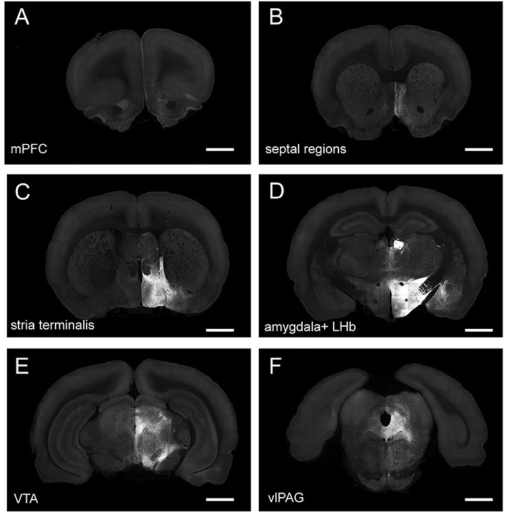
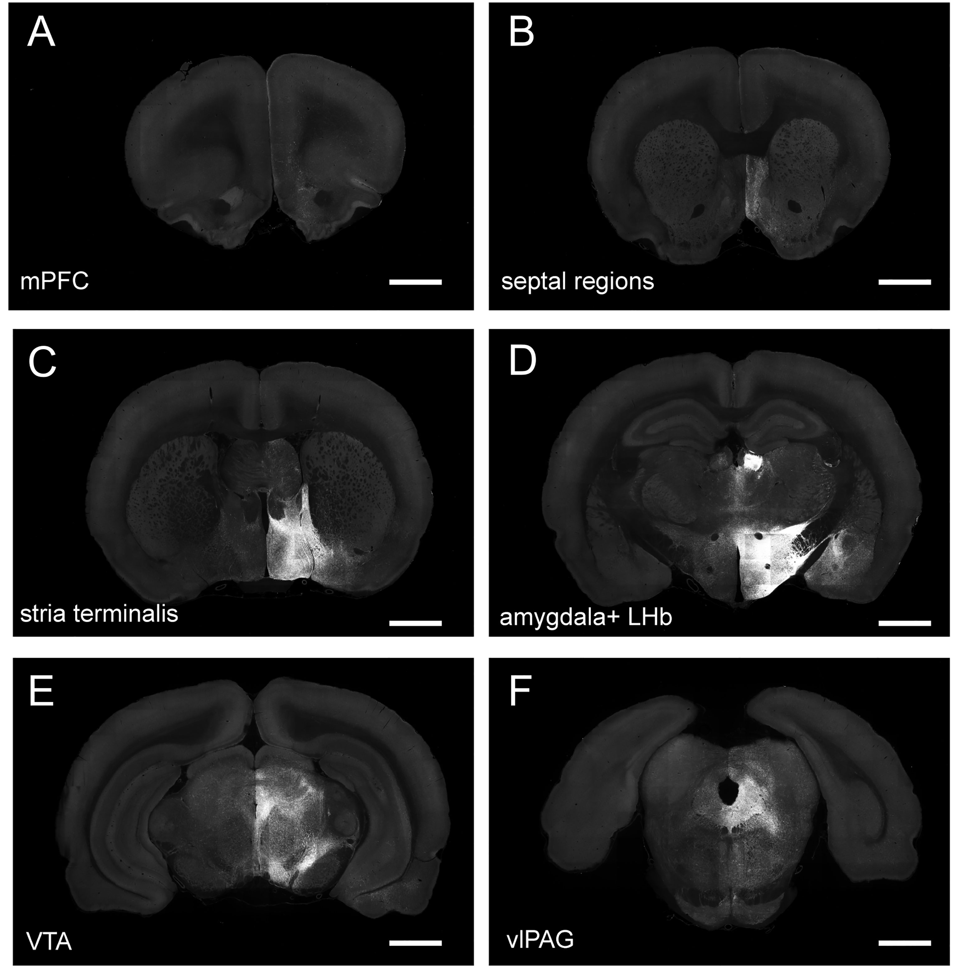
Figure 3:  Projections from virally transduced GABAergic neurons in lateral hypothalamus.  AAV1-EF1a-DIO-mem-AcGFP was injected into the LH, and brain tissue was imaged 2 weeks later using whole-brain TissueCyte system. There was intense membrane-GFP labeling in LH and lateral habenula and amygdala (D) and in the bed nucleus of the stria terminalis (C). Membrane-GFP labeling was also observed in VTA within the parabrachial pigmented area (E), septal regions (B), ventral-lateral periaqueductal gray (F), and lateral infralimbic (IL) and prelimbic (PL) cortices (mPFC; A). Views of stitched fields of coronal sections are shown. The scale bar represents 2 mm. Download print resolution version here.

Figures were modified and reprinted from “Sharpe et. al, Lateral Hypothalamic GABAergic Neurons Encode Reward Predictions that Are Relayed to the Ventral Tegmental Area to Regulate Learning. Curr Biol. 2017 Jul 24;27(14):2089-2100.e5. doi: 10.1016/j.cub.2017.06.024. Epub 2017 Jul 6” with permission from Elsevier.

For more details and data on phenotypic characterization, see reference (PMID: 28690111).

Breeding Strategy

Breeding Information, click here for PDF 

Genotyping Assays

Assay for the presence of Gad1-iCre transgene, click here for PDF

References that cite this rat

2022

Farrell, Mitchell R; Ye, Qiying; Xie, Yiyan; Esteban, Jeanine Sandra D; Mahler, Stephen V

Ventral pallidum GABA neurons bidirectionally control opioid relapse across rat behavioral models Journal Article

In: Addict Neurosci, vol. 3, 2022, ISSN: 2772-3925.

Abstract | Links

@article{pmid36156918,
title = {Ventral pallidum GABA neurons bidirectionally control opioid relapse across rat behavioral models},
author = {Mitchell R Farrell and Qiying Ye and Yiyan Xie and Jeanine Sandra D Esteban and Stephen V Mahler},
url = {https://pubmed.ncbi.nlm.nih.gov/36156918/},
doi = {10.1016/j.addicn.2022.100026},
issn = {2772-3925},
year = {2022},
date = {2022-09-01},
urldate = {2022-09-01},
journal = {Addict Neurosci},
volume = {3},
abstract = {Opioid addiction is a chronic, relapsing disorder. Whether addicted individuals are forced to abstain or they decide themselves to quit using drugs, relapse rates are high-especially upon encountering contexts and stimuli associated with prior opioid use. Rodents similarly show context- and cue-induced reinstatement of drug seeking following abstinence, and intriguingly, the neural circuits underlying these relapse-like behaviors differ when abstinence is involuntarily imposed, responding is extinguished, or animals decide themselves to cease taking drug. Here, we employ two complementary rat behavioral models of relapse-like behavior for the highly reinforcing opioid drug remifentanil, and asked whether GABAergic neurons in the ventral pallidum (VP) control opioid seeking under these behavioral conditions. Specifically, we asked how chemogenetically stimulating VP neurons with clozapine-N-oxide (CNO) influences the ability of contextual or discrete remifentanil-paired cues to reinstate drug seeking following either voluntary abstinence (punishment-induced; Group), or extinction training (Group). In Group rats, we also chemogenetically inhibited VP neurons, and examined spontaneous VP activity (Fos) during cued reinstatement. In both Group and Group rats, stimulating Gq-signaling in VP neurons augmented remifentanil reinstatement in a cue- and context-dependent manner. Conversely, engaging inhibitory Gi-signaling in VP neurons in Group suppressed cue-induced reinstatement, and cue-triggered seeking was correlated with Fos expression in rostral, but not caudal VP. Neither stimulating nor inhibiting VP neurons influenced unpunished remifentanil self-administration. We conclude that VP neurons bidirectionally control opioid seeking regardless of the specific relapse model employed, highlighting their fundamental role in opioid relapse-like behavior across behavioral models, and potentially across species.},
keywords = {},
pubstate = {published},
tppubtype = {article}
}

Close

Opioid addiction is a chronic, relapsing disorder. Whether addicted individuals are forced to abstain or they decide themselves to quit using drugs, relapse rates are high-especially upon encountering contexts and stimuli associated with prior opioid use. Rodents similarly show context- and cue-induced reinstatement of drug seeking following abstinence, and intriguingly, the neural circuits underlying these relapse-like behaviors differ when abstinence is involuntarily imposed, responding is extinguished, or animals decide themselves to cease taking drug. Here, we employ two complementary rat behavioral models of relapse-like behavior for the highly reinforcing opioid drug remifentanil, and asked whether GABAergic neurons in the ventral pallidum (VP) control opioid seeking under these behavioral conditions. Specifically, we asked how chemogenetically stimulating VP neurons with clozapine-N-oxide (CNO) influences the ability of contextual or discrete remifentanil-paired cues to reinstate drug seeking following either voluntary abstinence (punishment-induced; Group), or extinction training (Group). In Group rats, we also chemogenetically inhibited VP neurons, and examined spontaneous VP activity (Fos) during cued reinstatement. In both Group and Group rats, stimulating Gq-signaling in VP neurons augmented remifentanil reinstatement in a cue- and context-dependent manner. Conversely, engaging inhibitory Gi-signaling in VP neurons in Group suppressed cue-induced reinstatement, and cue-triggered seeking was correlated with Fos expression in rostral, but not caudal VP. Neither stimulating nor inhibiting VP neurons influenced unpunished remifentanil self-administration. We conclude that VP neurons bidirectionally control opioid seeking regardless of the specific relapse model employed, highlighting their fundamental role in opioid relapse-like behavior across behavioral models, and potentially across species.

Close

  • https://pubmed.ncbi.nlm.nih.gov/36156918/
  • doi:10.1016/j.addicn.2022.100026

Close

2020

Luo, Thomas Zhihao; Bondy, Adrian Gopnik; Gupta, Diksha; Elliott, Verity Alexander; Kopec, Charles D; Brody, Carlos D

An approach for long-term, multi-probe Neuropixels recordings in unrestrained rats Journal Article

In: eLife, vol. 9, pp. e59716, 2020, ISSN: 2050-084X.

Abstract | Links

@article{10.7554/eLife.59716,
title = {An approach for long-term, multi-probe Neuropixels recordings in unrestrained rats},
author = {Thomas Zhihao Luo and Adrian Gopnik Bondy and Diksha Gupta and Verity Alexander Elliott and Charles D Kopec and Carlos D Brody},
editor = {Lisa Giocomo and Laura L Colgin and Lisa Giocomo and Anne K Churchland},
url = {https://pubmed.ncbi.nlm.nih.gov/33089778/},
doi = {10.7554/eLife.59716},
issn = {2050-084X},
year = {2020},
date = {2020-10-01},
journal = {eLife},
volume = {9},
pages = {e59716},
publisher = {eLife Sciences Publications, Ltd},
abstract = {The use of Neuropixels probes for chronic neural recordings is in its infancy and initial studies leave questions about long-term stability and probe reusability unaddressed. Here, we demonstrate a new approach for chronic Neuropixels recordings over a period of months in freely moving rats. Our approach allows multiple probes per rat and multiple cycles of probe reuse. We found that hundreds of units could be recorded for multiple months, but that yields depended systematically on anatomical position. Explanted probes displayed a small increase in noise compared to unimplanted probes, but this was insufficient to impair future single-unit recordings. We conclude that cost-effective, multi-region, and multi-probe Neuropixels recordings can be carried out with high yields over multiple months in rats or other similarly sized animals. Our methods and observations may facilitate the standardization of chronic recording from Neuropixels probes in freely moving animals.},
keywords = {},
pubstate = {published},
tppubtype = {article}
}

Close

The use of Neuropixels probes for chronic neural recordings is in its infancy and initial studies leave questions about long-term stability and probe reusability unaddressed. Here, we demonstrate a new approach for chronic Neuropixels recordings over a period of months in freely moving rats. Our approach allows multiple probes per rat and multiple cycles of probe reuse. We found that hundreds of units could be recorded for multiple months, but that yields depended systematically on anatomical position. Explanted probes displayed a small increase in noise compared to unimplanted probes, but this was insufficient to impair future single-unit recordings. We conclude that cost-effective, multi-region, and multi-probe Neuropixels recordings can be carried out with high yields over multiple months in rats or other similarly sized animals. Our methods and observations may facilitate the standardization of chronic recording from Neuropixels probes in freely moving animals.

Close

  • https://pubmed.ncbi.nlm.nih.gov/33089778/
  • doi:10.7554/eLife.59716

Close

Prasad, Asheeta A; Xie, Caroline; Chaichim, Chanchanok; Nguyen, Jennifer H; McClusky, Hannah E; Killcross, Simon; Power, John M; McNally, Gavan P

Complementary Roles for Ventral Pallidum Cell Types and Their Projections in Relapse Journal Article

In: Journal of Neuroscience, vol. 40, no. 4, pp. 880–893, 2020, ISSN: 0270-6474.

Abstract | Links

@article{Prasad880,
title = {Complementary Roles for Ventral Pallidum Cell Types and Their Projections in Relapse},
author = {Asheeta A Prasad and Caroline Xie and Chanchanok Chaichim and Jennifer H Nguyen and Hannah E McClusky and Simon Killcross and John M Power and Gavan P McNally},
url = {https://pubmed.ncbi.nlm.nih.gov/31818977/},
doi = {10.1523/JNEUROSCI.0262-19.2019},
issn = {0270-6474},
year = {2020},
date = {2020-01-01},
journal = {Journal of Neuroscience},
volume = {40},
number = {4},
pages = {880--893},
publisher = {Society for Neuroscience},
abstract = {The ventral pallidum (VP) is a key node in the neural circuits controlling relapse to drug seeking. How this role relates to different VP cell types and their projections is poorly understood. Using male rats, we show how different forms of relapse to alcohol-seeking are assembled from VP cell types and their projections to lateral hypothalamus (LH) and ventral tegmental area (VTA). Using RNAScope in situ hybridization to characterize activity of different VP cell types during relapse to alcohol-seeking provoked by renewal (context-induced reinstatement), we found that VP Gad1 and parvalbumin (PV), but not vGlut2, neurons show relapse-associated changes in c-Fos expression. Next, we used retrograde tracing, chemogenetic, and electrophysiological approaches to study the roles of VPGad1 and VPPV neurons in relapse. We show that VPGad1 neurons contribute to contextual control over relapse (renewal), but not to relapse during reacquisition, via projections to LH, where they converge with ventral striatal inputs onto LHGad1 neurons. This convergence of striatopallidal inputs at the level of individual LHGad1 neurons may be critical to balancing propensity for relapse versus abstinence. In contrast, VPPV neurons contribute to relapse during both renewal and reacquisition via projections to VTA. These findings identify a double dissociation in the roles for different VP cell types and their projections in relapse. VPGad1 neurons control relapse during renewal via projections to LH. VPPV neurons control relapse during both renewal and reacquisition via projections to VTA. Targeting these different pathways may provide tailored interventions for different forms of relapse.SIGNIFICANCE STATEMENT Relapse to drug or reward seeking after a period of extinction or abstinence remains a key impediment to successful treatment. The ventral pallidum, located in the ventral basal ganglia, has long been recognized as an obligatory node in a textquoterightfinal common pathwaytextquoteright for relapse. Yet how this role relates to the considerable VP cellular and circuit heterogeneity is not well understood. We studied the cellular and circuit architecture for VP in relapse control. We show that different forms of relapse have complementary VP cellular and circuit architectures, raising the possibility that targeting these different neural architectures may provide tailored interventions for different forms of relapse.},
keywords = {},
pubstate = {published},
tppubtype = {article}
}

Close

The ventral pallidum (VP) is a key node in the neural circuits controlling relapse to drug seeking. How this role relates to different VP cell types and their projections is poorly understood. Using male rats, we show how different forms of relapse to alcohol-seeking are assembled from VP cell types and their projections to lateral hypothalamus (LH) and ventral tegmental area (VTA). Using RNAScope in situ hybridization to characterize activity of different VP cell types during relapse to alcohol-seeking provoked by renewal (context-induced reinstatement), we found that VP Gad1 and parvalbumin (PV), but not vGlut2, neurons show relapse-associated changes in c-Fos expression. Next, we used retrograde tracing, chemogenetic, and electrophysiological approaches to study the roles of VPGad1 and VPPV neurons in relapse. We show that VPGad1 neurons contribute to contextual control over relapse (renewal), but not to relapse during reacquisition, via projections to LH, where they converge with ventral striatal inputs onto LHGad1 neurons. This convergence of striatopallidal inputs at the level of individual LHGad1 neurons may be critical to balancing propensity for relapse versus abstinence. In contrast, VPPV neurons contribute to relapse during both renewal and reacquisition via projections to VTA. These findings identify a double dissociation in the roles for different VP cell types and their projections in relapse. VPGad1 neurons control relapse during renewal via projections to LH. VPPV neurons control relapse during both renewal and reacquisition via projections to VTA. Targeting these different pathways may provide tailored interventions for different forms of relapse.SIGNIFICANCE STATEMENT Relapse to drug or reward seeking after a period of extinction or abstinence remains a key impediment to successful treatment. The ventral pallidum, located in the ventral basal ganglia, has long been recognized as an obligatory node in a textquoterightfinal common pathwaytextquoteright for relapse. Yet how this role relates to the considerable VP cellular and circuit heterogeneity is not well understood. We studied the cellular and circuit architecture for VP in relapse control. We show that different forms of relapse have complementary VP cellular and circuit architectures, raising the possibility that targeting these different neural architectures may provide tailored interventions for different forms of relapse.

Close

  • https://pubmed.ncbi.nlm.nih.gov/31818977/
  • doi:10.1523/JNEUROSCI.0262-19.2019

Close

2018

Gibson, Gabrielle D; Prasad, Asheeta A; Jean-Richard-dit-Bressel, Philip; Yau, Joanna O Y; Millan, Zayra E; Liu, Yu; Campbell, Erin J; Lim, Jun; Marchant, Nathan J; Power, John M; Killcross, Simon; Lawrence, Andrew J; McNally, Gavan P

Distinct Accumbens Shell Output Pathways Promote versus Prevent Relapse to Alcohol Seeking Journal Article

In: Neuron, vol. 98, no. 3, pp. 512–520.e6, 2018, ISBN: 0896-6273.

Abstract | Links

@article{Gibson:2018aa,
title = {Distinct Accumbens Shell Output Pathways Promote versus Prevent Relapse to Alcohol Seeking},
author = {Gabrielle D Gibson and Asheeta A Prasad and Philip Jean-Richard-dit-Bressel and Joanna O Y Yau and Zayra E Millan and Yu Liu and Erin J Campbell and Jun Lim and Nathan J Marchant and John M Power and Simon Killcross and Andrew J Lawrence and Gavan P McNally},
url = {https://pubmed.ncbi.nlm.nih.gov/29656870/},
doi = {10.1016/j.neuron.2018.03.033},
isbn = {0896-6273},
year = {2018},
date = {2018-05-02},
booktitle = {Neuron},
journal = {Neuron},
volume = {98},
number = {3},
pages = {512--520.e6},
publisher = {Elsevier},
abstract = {Contexts exert bi-directional control over relapse to drug seeking. Contexts associated with drug self-administration promote relapse, whereas contexts associated with the absence of self-administration protect against relapse. The nucleus accumbens shell (AcbSh) is a key brain region determining these roles of context. However, the specific cell types, and projections, by which AcbSh serves these dual roles are unknown. Here, we show that contextual control over relapse and abstinence is embedded within distinct output circuits of dopamine 1 receptor (Drd1) expressing AcbSh neurons. We report anatomical and functional segregation of Drd1 AcbSh output pathways during context-induced reinstatement and extinction of alcohol seeking. The AcbSh?ventral tegmental area (VTA) pathway promotes relapse via projections to VTA Gad1 neurons. The AcbSh?lateral hypothalamus (LH) pathway promotes extinction via projections to LH Gad1 neurons. Targeting these opposing AcbSh circuit contributions may reduce propensity to relapse to, and promote abstinence from, drug use.},
keywords = {},
pubstate = {published},
tppubtype = {article}
}

Close

Contexts exert bi-directional control over relapse to drug seeking. Contexts associated with drug self-administration promote relapse, whereas contexts associated with the absence of self-administration protect against relapse. The nucleus accumbens shell (AcbSh) is a key brain region determining these roles of context. However, the specific cell types, and projections, by which AcbSh serves these dual roles are unknown. Here, we show that contextual control over relapse and abstinence is embedded within distinct output circuits of dopamine 1 receptor (Drd1) expressing AcbSh neurons. We report anatomical and functional segregation of Drd1 AcbSh output pathways during context-induced reinstatement and extinction of alcohol seeking. The AcbSh?ventral tegmental area (VTA) pathway promotes relapse via projections to VTA Gad1 neurons. The AcbSh?lateral hypothalamus (LH) pathway promotes extinction via projections to LH Gad1 neurons. Targeting these opposing AcbSh circuit contributions may reduce propensity to relapse to, and promote abstinence from, drug use.

Close

  • https://pubmed.ncbi.nlm.nih.gov/29656870/
  • doi:10.1016/j.neuron.2018.03.033

Close

2017

Sharpe, Melissa J; Marchant, Nathan J; Whitaker, Leslie R; Richie, Christopher T; Zhang, Yajun J; Campbell, Erin J; Koivula, Pyry P; Necarsulmer, Julie C; Mejias-Aponte, Carlos; Morales, Marisela; Pickel, James; Smith, Jeffrey C; Niv, Yael; Shaham, Yavin; Harvey, Brandon K; Schoenbaum, Geoffrey

Lateral Hypothalamic GABAergic Neurons Encode Reward Predictions that Are Relayed to the Ventral Tegmental Area to Regulate Learning. Journal Article

In: Curr Biol, vol. 27, no. 14, pp. 2089–2100, 2017, ISSN: 1879-0445 (Electronic); 0960-9822 (Linking), (*First paper describing LE-Tg(GAD1-iCre)3Ottc rat.).

Abstract | Links

@article{Sharpe:2017aab,
title = {Lateral Hypothalamic GABAergic Neurons Encode Reward Predictions that Are Relayed to the Ventral Tegmental Area to Regulate Learning.},
author = {Melissa J Sharpe and Nathan J Marchant and Leslie R Whitaker and Christopher T Richie and Yajun J Zhang and Erin J Campbell and Pyry P Koivula and Julie C Necarsulmer and Carlos Mejias-Aponte and Marisela Morales and James Pickel and Jeffrey C Smith and Yael Niv and Yavin Shaham and Brandon K Harvey and Geoffrey Schoenbaum},
url = {https://www.ncbi.nlm.nih.gov/pubmed/28690111},
doi = {10.1016/j.cub.2017.06.024},
issn = {1879-0445 (Electronic); 0960-9822 (Linking)},
year = {2017},
date = {2017-07-06},
urldate = {2017-07-06},
journal = {Curr Biol},
volume = {27},
number = {14},
pages = {2089--2100},
address = {National Institute on Drug Abuse, IRP, 251 Bayview Boulevard, Baltimore, MD 21228, USA; Princeton Neuroscience Institute, Princeton University, Washington Road, Princeton, NJ 08544, USA. Electronic address: melissa.sharpe@nih.gov.},
abstract = {Eating is a learned process. Our desires for specific foods arise through experience. Both electrical stimulation and optogenetic studies have shown that increased activity in the lateral hypothalamus (LH) promotes feeding. Current dogma is that these effects reflect a role for LH neurons in the control of the core motivation to feed, and their activity comes under control of forebrain regions to elicit learned food-motivated behaviors. However, these effects could also reflect the storage of associative information about the cues leading to food in LH itself. Here, we present data from several studies that are consistent with a role for LH in learning. In the first experiment, we use a novel GAD-Cre rat to show that optogenetic inhibition of LH gamma-aminobutyric acid (GABA) neurons restricted to cue presentation disrupts the rats' ability to learn that a cue predicts food without affecting subsequent food consumption. In the second experiment, we show that this manipulation also disrupts the ability of a cue to promote food seeking after learning. Finally, we show that inhibition of the terminals of the LH GABA neurons in ventral-tegmental area (VTA) facilitates learning about reward-paired cues. These results suggest that the LH GABA neurons are critical for storing and later disseminating information about reward-predictive cues.},
note = {*First paper describing LE-Tg(GAD1-iCre)3Ottc rat.},
keywords = {},
pubstate = {published},
tppubtype = {article}
}

Close

Eating is a learned process. Our desires for specific foods arise through experience. Both electrical stimulation and optogenetic studies have shown that increased activity in the lateral hypothalamus (LH) promotes feeding. Current dogma is that these effects reflect a role for LH neurons in the control of the core motivation to feed, and their activity comes under control of forebrain regions to elicit learned food-motivated behaviors. However, these effects could also reflect the storage of associative information about the cues leading to food in LH itself. Here, we present data from several studies that are consistent with a role for LH in learning. In the first experiment, we use a novel GAD-Cre rat to show that optogenetic inhibition of LH gamma-aminobutyric acid (GABA) neurons restricted to cue presentation disrupts the rats' ability to learn that a cue predicts food without affecting subsequent food consumption. In the second experiment, we show that this manipulation also disrupts the ability of a cue to promote food seeking after learning. Finally, we show that inhibition of the terminals of the LH GABA neurons in ventral-tegmental area (VTA) facilitates learning about reward-paired cues. These results suggest that the LH GABA neurons are critical for storing and later disseminating information about reward-predictive cues.

Close

  • https://www.ncbi.nlm.nih.gov/pubmed/28690111
  • doi:10.1016/j.cub.2017.06.024

Close

Blog/Comments/Reviews

Last Updated on November 12, 2024

There are 26 surveyed reports for the receiving and usage of the transgenic GAD1-iCre rats with only 3 total reported issues.

General Health
One lab group noticed patches of fur loss at 4 months of age in rats that was resolved in about 2 weeks.
Another lab group discovered a large tumor in one of the original GAD1-iCre breeders.

Weight
There are no reports of weight issues with the LE-Tg(GAD-iCre)3Ottc rats

Breeding
There are no reports of breeding issues with the LE-Tg(GAD1-iCre)3Ottc rats

Expression
One lab group experienced variable Cre expression with inconsistencies in Cre and GAD1 labeled positive neurons when examining the nucleus accumbens core, ventral pallidum, and dorsal raphe brain regions

*Please be sure to follow the breeding guide to produce a working LE-Tg(Drd1a-iCre)3Ottc colony.


Other related rats

No related rats at this time.

Acknowledgements

YaJun Zhang, Julie Necarsulmer, Chris Richie, Brandon Harvey, Janette Lebron, Melissa Sharpe, Geoffrey Schoenbaum


Primary Sidebar

Transgenic Rats Project

  • Transgenic Rat Home
  • OTTC Information
  • Publications
  • Technology Development Initiative
  • Links

Organization

  • Organization
  • Faculty
  • Office of the Scientific Director
  • Office of the Clinical Director
  • Administrative Management Branch
  • Molecular Targets and Medications Discovery Branch
  • Cellular and Neurocomputational Systems Branch
  • Molecular Neuropsychiatry Research Branch
  • Neuroimaging Research Branch
  • Behavioral Neuroscience Research Branch
  • Integrative Neuroscience Research Branch
  • Translational Addiction Medicine Branch
  • Core Facilities
  • Careers at NIDA IRP
  • Technology Development Initiative
  • Community Outreach Group
Home / Organization / Office of the Scientific Director / Transgenic Rat Project / LE-Tg(Gad1-iCre)3Ottc
  • National Institute on Drug Abuse
  • NIH Intramural Research Program
  • National Institutes of Health
  • Health and Human Services
  • USA.GOV
  • Emergency Contacts
  • Employee Assistance
  • Treatment Information
  • Contact Us
  • Careers at NIDA IRP
  • Accessibility
  • Privacy
  • HHS Vulnerability Disclosure
  • Freedom of Information Act
  • Document Viewing Tools
  • Offsite Links

  • Home
  • News
    ▼
    • Featured Paper of the Month
    • Reviews to Read
    • Hot off the Press
    • IRP News
    • Awards
    • Technology Development Initiative Paper of the Month
    • Seminar Series
    • Addiction Grand Rounds
  • About
    ▼
    • About NIDA IRP
    • Contact Us
    • Directions and Map
    • Careers at NIDA IRP
    • Emergency Contacts
    • Employee Assistance Resources
  • Organization
    ▼
    • Faculty
    • Office of the Scientific Director
    • Office of the Clinical Director
    • Office of Education and Career Development
    • Administrative Management Branch
    • Molecular Targets and Medications Discovery Branch
    • Cellular and Neurocomputational Systems Branch
    • Molecular Neuropsychiatry Research Branch
    • Neuroimaging Research Branch
    • Behavioral Neuroscience Research Branch
    • Integrative Neuroscience Research Branch
    • Translational Addiction Medicine Branch
    • Core Facilities
    • Community Outreach Group
  • Training Programs
    ▼
    • Office of Education and Career Development
    • OECD Awards
    • Summer Internship Program
    • Postbaccalaureate Program
    • Graduate Partnership Program
    • Postdoctoral Program
    • NIDA Speakers Bureau
    • Clinical Electives Program
    • Clinical Mentoring Program
  • Study Volunteers