• Skip to primary navigation
  • Skip to main content
  • Skip to primary sidebar

NIDA IRP

National Institute on Drug Abuse - Intramural Research Program

  National Institute on Drug Abuse | NIH IRP | Treatment Info | Emergency Contacts
  • Home
  • News
    • Featured Paper of the Month
    • Reviews to Read
    • Hot off the Press
    • IRP News
    • Awards
    • Technology Development Initiative Paper of the Month
    • Seminar Series
    • Addiction Grand Rounds
  • About
    • About NIDA IRP
    • Contact Us
    • Directions and Map
    • Careers at NIDA IRP
    • Emergency Contacts
    • Employee Assistance Resources
  • Organization
    • Faculty
    • Office of the Scientific Director
    • Office of the Clinical Director
    • Office of Education and Career Development
    • Administrative Management Branch
    • Molecular Targets and Medications Discovery Branch
    • Cellular and Neurocomputational Systems Branch
    • Molecular Neuropsychiatry Research Branch
    • Neuroimaging Research Branch
    • Behavioral Neuroscience Research Branch
    • Integrative Neuroscience Research Branch
    • Translational Addiction Medicine Branch
    • Core Facilities
    • Community Outreach Group
  • Training Programs
    • Office of Education and Career Development
    • OECD Awards
    • Summer Internship Program
    • Postbaccalaureate Program
    • Graduate Partnership Program
    • Postdoctoral Program
    • NIDA Speakers Bureau
    • Clinical Electives Program
    • Clinical Mentoring Program
  • Study Volunteers
  • Transgenic Rat Home
  • OTTC Information
  • Publications
  • Technology Development Initiative
  • Links

LE-Tg(Drd2-iCre)1Ottc

Last Updated on November 12, 2024

Background | Status & Availability | Transgene Info | Phenotypic Characterization | Breeding | Genotyping | References | Blog/Comments/Reviews | Related rats | Acknowledgements

Background

The dopamine receptor Drd2 is highly expressed in medium spiny neurons (MSNs) of the  striatum.   The Drd2-expressing  MSNs send inhibitory projections to the globus pallidus externa (GPe) which in turn sends inhibitory projections to the sub-thalamic nucleus (STN).  The STN neurons have excitory projections that activate basal ganglia structures such as the globus pallidus interna (GPi) and substantia nigra reticulata (SNr).  This collection of projecting  neurons is referred to as the “indirect pathway” or “D2 pathway”.   The indirect pathway is important for controlling voluntary movements and reward-related behaviors. We have generated and characterized a strain of transgenic Long Evans rats expressing iCre recombinase under a Drd2 promoter referred to as “LE-Tg(Drd2-iCre)1Ottc.”  The tissue-specific expression of iCre can be used in combination with Cre-dependent transgenes to obtain selective expression of transgenes in Drd2(+) cells.

Status and Availability

This strain has been published (PMID: 33051346).
As of February 21, 2017, this strain is available as line 768 at the RRRC.
This rat is registered at the Rat Genome Database (RGD) as RGD ID#10412327.

Transgene Information

Schematic Draft - Drd2-iCre

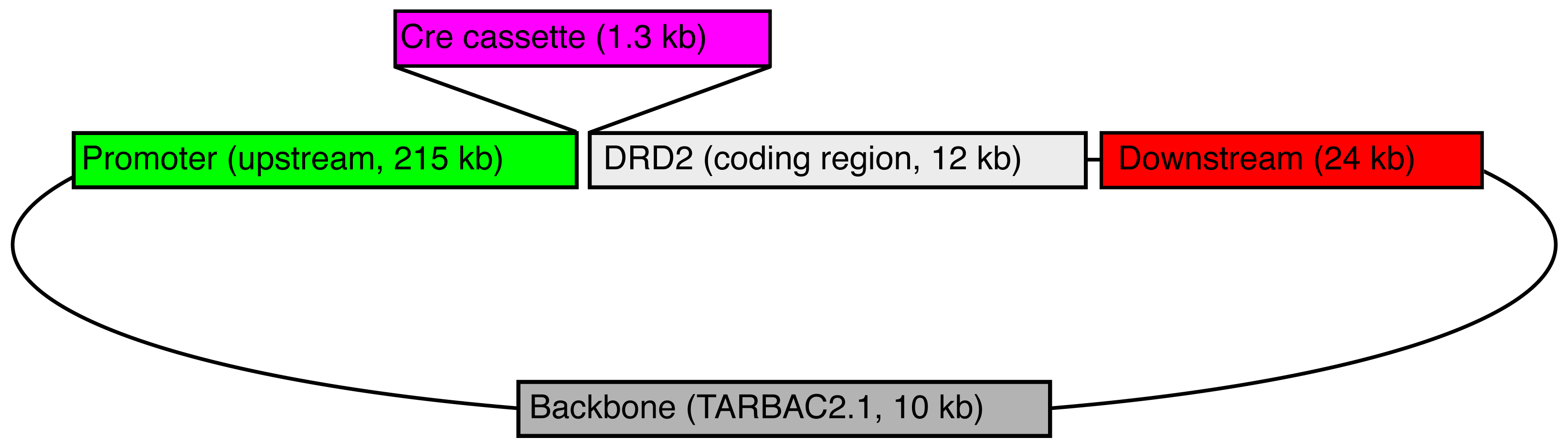
Legend:  A bacterial artificial chromosome (BAC) containing the rat Drd2 gene (CH230-11B15) was obtained from CHORI, and recombineered to replace the start codon of Drd2 with a 1.3 kb cassette containing iCre (improved Cre recombinase), the polyadenylation signal from the gene for bovine growth hormone, and a galk bacterial selection marker (pOTTC464).  This BAC was injected into the pronuclei of fertilized Long Evans rat embryos by NIMH Transgenic Core, and ultimately resulted in two independent, phenotypically positive Drd2-iCre lines.  This line (LE-Tg(Drd2-iCre)1Ottc) has four copies of the transgene per haploid genome as determined by droplet digital PCR.

Phenotypic Characterization

To be added.  Please check back.

Breeding Strategy

Breeding Information, click here for PDF

Genotyping Assays

Assay for the presence of Drd2 and iCre, click here for PDF

References that cite this rat

2023

Truckenbrod, Leah M; Betzhold, Sara M; Wheeler, Alexa-Rae; Shallcross, John; Singhal, Sarthak; Harden, Scott; Schwendt, Marek; Frazier, Charles J; Bizon, Jennifer L; Setlow, Barry; Orsini, Caitlin A

Circuit and Cell-Specific Contributions to Decision Making Involving Risk of Explicit Punishment in Male and Female Rats Journal Article

In: J Neurosci, vol. 43, no. 26, pp. 4837–4855, 2023, ISSN: 1529-2401.

Abstract | Links

@article{pmid37286352,
title = {Circuit and Cell-Specific Contributions to Decision Making Involving Risk of Explicit Punishment in Male and Female Rats},
author = {Leah M Truckenbrod and Sara M Betzhold and Alexa-Rae Wheeler and John Shallcross and Sarthak Singhal and Scott Harden and Marek Schwendt and Charles J Frazier and Jennifer L Bizon and Barry Setlow and Caitlin A Orsini},
url = {https://pubmed.ncbi.nlm.nih.gov/37286352/},
doi = {10.1523/JNEUROSCI.0276-23.2023},
issn = {1529-2401},
year = {2023},
date = {2023-06-01},
urldate = {2023-06-01},
journal = {J Neurosci},
volume = {43},
number = {26},
pages = {4837--4855},
abstract = {Decision making is a complex cognitive process that recruits a distributed network of brain regions, including the basolateral amygdala (BLA) and nucleus accumbens shell (NAcSh). Recent work suggests that communication between these structures, as well as activity of cells expressing dopamine (DA) D2 receptors (D2R) in the NAcSh, are necessary for some forms of decision making; however, the contributions of this circuit and cell population during decision making under risk of punishment are unknown. The current experiments addressed this question using circuit-specific and cell type-specific optogenetic approaches in rats during a decision making task involving risk of punishment. In experiment 1, Long-Evans rats received intra-BLA injections of halorhodopsin or mCherry (control) and in experiment 2, D2-Cre transgenic rats received intra-NAcSh injections of Cre-dependent halorhodopsin or mCherry. Optic fibers were implanted in the NAcSh in both experiments. Following training in the decision making task, BLA→NAcSh or D2R-expressing neurons were optogenetically inhibited during different phases of the decision process. Inhibition of the BLA→NAcSh during deliberation (the time between trial initiation and choice) increased preference for the large, risky reward (increased risk taking). Similarly, inhibition during delivery of the large, punished reward increased risk taking, but only in males. Inhibition of D2R-expressing neurons in the NAcSh during deliberation increased risk taking. In contrast, inhibition of these neurons during delivery of the small, safe reward decreased risk taking. These findings extend our knowledge of the neural dynamics of risk taking, revealing sex-dependent circuit recruitment and dissociable activity of selective cell populations during decision making. Until recently, the ability to dissect the neural substrates of decision making involving risk of punishment (risk taking) in a circuit-specific and cell-specific manner has been limited by the tools available for use in rats. Here, we leveraged the temporal precision of optogenetics, together with transgenic rats, to probe contributions of a specific circuit and cell population to different phases of risk-based decision making. Our findings reveal basolateral amygdala (BLA)→nucleus accumbens shell (NAcSh) is involved in evaluation of punished rewards in a sex-dependent manner. Further, NAcSh D2 receptor (D2R)-expressing neurons make unique contributions to risk taking that vary across the decision making process. These findings advance our understanding of the neural principles of decision making and provide insight into how risk taking may become compromised in neuropsychiatric diseases.},
keywords = {},
pubstate = {published},
tppubtype = {article}
}

Close

Decision making is a complex cognitive process that recruits a distributed network of brain regions, including the basolateral amygdala (BLA) and nucleus accumbens shell (NAcSh). Recent work suggests that communication between these structures, as well as activity of cells expressing dopamine (DA) D2 receptors (D2R) in the NAcSh, are necessary for some forms of decision making; however, the contributions of this circuit and cell population during decision making under risk of punishment are unknown. The current experiments addressed this question using circuit-specific and cell type-specific optogenetic approaches in rats during a decision making task involving risk of punishment. In experiment 1, Long-Evans rats received intra-BLA injections of halorhodopsin or mCherry (control) and in experiment 2, D2-Cre transgenic rats received intra-NAcSh injections of Cre-dependent halorhodopsin or mCherry. Optic fibers were implanted in the NAcSh in both experiments. Following training in the decision making task, BLA→NAcSh or D2R-expressing neurons were optogenetically inhibited during different phases of the decision process. Inhibition of the BLA→NAcSh during deliberation (the time between trial initiation and choice) increased preference for the large, risky reward (increased risk taking). Similarly, inhibition during delivery of the large, punished reward increased risk taking, but only in males. Inhibition of D2R-expressing neurons in the NAcSh during deliberation increased risk taking. In contrast, inhibition of these neurons during delivery of the small, safe reward decreased risk taking. These findings extend our knowledge of the neural dynamics of risk taking, revealing sex-dependent circuit recruitment and dissociable activity of selective cell populations during decision making. Until recently, the ability to dissect the neural substrates of decision making involving risk of punishment (risk taking) in a circuit-specific and cell-specific manner has been limited by the tools available for use in rats. Here, we leveraged the temporal precision of optogenetics, together with transgenic rats, to probe contributions of a specific circuit and cell population to different phases of risk-based decision making. Our findings reveal basolateral amygdala (BLA)→nucleus accumbens shell (NAcSh) is involved in evaluation of punished rewards in a sex-dependent manner. Further, NAcSh D2 receptor (D2R)-expressing neurons make unique contributions to risk taking that vary across the decision making process. These findings advance our understanding of the neural principles of decision making and provide insight into how risk taking may become compromised in neuropsychiatric diseases.

Close

  • https://pubmed.ncbi.nlm.nih.gov/37286352/
  • doi:10.1523/JNEUROSCI.0276-23.2023

Close

Swinford-Jackson, Sarah E; Huffman, Phillip J; Knouse, Melissa C; Thomas, Arthur S; Rich, Matthew T; Mankame, Sharvari; Worobey, Samantha J; Sarmiento, Mateo; Coleman, Ayanna; Pierce, R Christopher

High frequency DBS-like optogenetic stimulation of nucleus accumbens dopamine D2 receptor-containing neurons attenuates cocaine reinstatement in male rats Journal Article

In: Neuropsychopharmacology, vol. 48, no. 3, pp. 459–467, 2023, ISSN: 1740-634X.

Abstract | Links

@article{pmid36446928,
title = {High frequency DBS-like optogenetic stimulation of nucleus accumbens dopamine D2 receptor-containing neurons attenuates cocaine reinstatement in male rats},
author = {Sarah E Swinford-Jackson and Phillip J Huffman and Melissa C Knouse and Arthur S Thomas and Matthew T Rich and Sharvari Mankame and Samantha J Worobey and Mateo Sarmiento and Ayanna Coleman and R Christopher Pierce},
url = {https://pubmed.ncbi.nlm.nih.gov/36446928/},
doi = {10.1038/s41386-022-01495-y},
issn = {1740-634X},
year = {2023},
date = {2023-02-01},
urldate = {2023-02-01},
journal = {Neuropsychopharmacology},
volume = {48},
number = {3},
pages = {459--467},
abstract = {Previous work indicated that deep brain stimulation (DBS) of the nucleus accumbens shell in male rats attenuated reinstatement of cocaine seeking, an animal model of craving. However, the potential differential impact of DBS on specific populations of neurons to drive the suppression of cocaine seeking is unknown. Medium spiny neurons in the nucleus accumbens are differentiated by expression of dopamine D1 receptors (D1DRs) or D2DRs, activation of which promotes or inhibits cocaine-related behaviors, respectively. The advent of transgenic rat lines expressing Cre recombinase selectively in D1DR-containing or D2DR-containing neurons, when coupled with Cre-dependent virally mediated gene transfer of channelrhodopsin (ChR2), enabled mimicry of DBS in a selective subpopulation of neurons during complex tasks. We tested the hypothesis that high frequency DBS-like optogenetic stimulation of D1DR-containing neurons in the accumbens shell would potentiate, whereas stimulation of D2DR-containing neurons in the accumbens shell would attenuate, cocaine-primed reinstatement of cocaine seeking. Results indicated that high frequency, DBS-like optogenetic stimulation of D2DR-containing neurons attenuated reinstatement of cocaine seeking in male rats, whereas DBS-like stimulation of D1DR-containing neurons did not alter cocaine-primed reinstatement. Surprisingly, DBS-like optogenetic stimulation did not alter reinstatement of cocaine seeking in female rats. In rats which only expressed eYFP, intra-accumbens optogenetic stimulation did not alter cocaine reinstatement, indicating that the effect of DBS-like stimulation to attenuate cocaine reinstatement is mediated specifically by ChR2 rather than by prolonged light delivery. These results suggest that DBS of the accumbens may attenuate cocaine-primed reinstatement in male rats through the selective manipulation of D2DR-containing neurons.},
keywords = {},
pubstate = {published},
tppubtype = {article}
}

Close

Previous work indicated that deep brain stimulation (DBS) of the nucleus accumbens shell in male rats attenuated reinstatement of cocaine seeking, an animal model of craving. However, the potential differential impact of DBS on specific populations of neurons to drive the suppression of cocaine seeking is unknown. Medium spiny neurons in the nucleus accumbens are differentiated by expression of dopamine D1 receptors (D1DRs) or D2DRs, activation of which promotes or inhibits cocaine-related behaviors, respectively. The advent of transgenic rat lines expressing Cre recombinase selectively in D1DR-containing or D2DR-containing neurons, when coupled with Cre-dependent virally mediated gene transfer of channelrhodopsin (ChR2), enabled mimicry of DBS in a selective subpopulation of neurons during complex tasks. We tested the hypothesis that high frequency DBS-like optogenetic stimulation of D1DR-containing neurons in the accumbens shell would potentiate, whereas stimulation of D2DR-containing neurons in the accumbens shell would attenuate, cocaine-primed reinstatement of cocaine seeking. Results indicated that high frequency, DBS-like optogenetic stimulation of D2DR-containing neurons attenuated reinstatement of cocaine seeking in male rats, whereas DBS-like stimulation of D1DR-containing neurons did not alter cocaine-primed reinstatement. Surprisingly, DBS-like optogenetic stimulation did not alter reinstatement of cocaine seeking in female rats. In rats which only expressed eYFP, intra-accumbens optogenetic stimulation did not alter cocaine reinstatement, indicating that the effect of DBS-like stimulation to attenuate cocaine reinstatement is mediated specifically by ChR2 rather than by prolonged light delivery. These results suggest that DBS of the accumbens may attenuate cocaine-primed reinstatement in male rats through the selective manipulation of D2DR-containing neurons.

Close

  • https://pubmed.ncbi.nlm.nih.gov/36446928/
  • doi:10.1038/s41386-022-01495-y

Close

2020

Strong, C. E.; Hagarty, D. P.; Guerrero, A. Brea; Schoepfer, K. J.; Cajuste, S. M.; Kabbaj, M.

Chemogenetic selective manipulation of nucleus accumbens medium spiny neurons bidirectionally controls alcohol intake in male and female rats Journal Article

In: Scientific Reports, vol. 10, no. 1, pp. 19178, 2020, ISBN: 2045-2322.

Abstract | Links

@article{Strong:2020tx,
title = {Chemogenetic selective manipulation of nucleus accumbens medium spiny neurons bidirectionally controls alcohol intake in male and female rats},
author = {C. E. Strong and D. P. Hagarty and A. Brea Guerrero and K. J. Schoepfer and S. M. Cajuste and M. Kabbaj},
url = {https://pubmed.ncbi.nlm.nih.gov/33154463/},
doi = {10.1038/s41598-020-76183-2},
isbn = {2045-2322},
year = {2020},
date = {2020-11-05},
urldate = {2020-11-05},
journal = {Scientific Reports},
volume = {10},
number = {1},
pages = {19178},
abstract = {The nucleus accumbens (NAc), considered the hub of reward circuitry, is comprised of two medium spiny neuron (MSN) subtypes that are classified by their enrichment of dopamine 1 (D1) or 2 (D2) receptors. While reports indicate that alcohol increases excitatory neurotransmission exclusively on NAc D1-MSNs in male rats, it remains unknown how NAc MSNs control alcohol intake in either sex. Therefore, this study investigated how NAc MSNs mediate alcohol intake by using Drd1a-iCre and Drd2-iCre transgenic rats of both sexes. Intra-NAc infusions of Cre-inducible viral vectors containing stimulatory (hM3Dq) or inhibitory (hM4Di) designer receptors exclusively activated by designer drugs (DREADDs) were delivered after 4-weeks of alcohol intake, and clozapine-N-oxide (CNO) was administered to selectively manipulate NAc MSNs. Our results show that activation of NAc D1-MSNs increased alcohol intake 1-, 4-, and 24-h after the start of drinking while inhibition decreased it 1-h after the start of drinking, with no sex differences observed at any time point. Activation of NAc D2-MSNs had no impact on alcohol intake while inhibition increased alcohol intake in Drd2-iCre rats for 1-h in males and 4-h in females. These findings suggest opposing roles for how NAc D1- and D2-MSNs modulate alcohol intake in rats of both sexes.},
keywords = {},
pubstate = {published},
tppubtype = {article}
}

Close

The nucleus accumbens (NAc), considered the hub of reward circuitry, is comprised of two medium spiny neuron (MSN) subtypes that are classified by their enrichment of dopamine 1 (D1) or 2 (D2) receptors. While reports indicate that alcohol increases excitatory neurotransmission exclusively on NAc D1-MSNs in male rats, it remains unknown how NAc MSNs control alcohol intake in either sex. Therefore, this study investigated how NAc MSNs mediate alcohol intake by using Drd1a-iCre and Drd2-iCre transgenic rats of both sexes. Intra-NAc infusions of Cre-inducible viral vectors containing stimulatory (hM3Dq) or inhibitory (hM4Di) designer receptors exclusively activated by designer drugs (DREADDs) were delivered after 4-weeks of alcohol intake, and clozapine-N-oxide (CNO) was administered to selectively manipulate NAc MSNs. Our results show that activation of NAc D1-MSNs increased alcohol intake 1-, 4-, and 24-h after the start of drinking while inhibition decreased it 1-h after the start of drinking, with no sex differences observed at any time point. Activation of NAc D2-MSNs had no impact on alcohol intake while inhibition increased alcohol intake in Drd2-iCre rats for 1-h in males and 4-h in females. These findings suggest opposing roles for how NAc D1- and D2-MSNs modulate alcohol intake in rats of both sexes.

Close

  • https://pubmed.ncbi.nlm.nih.gov/33154463/
  • doi:10.1038/s41598-020-76183-2

Close

Luo, Thomas Zhihao; Bondy, Adrian Gopnik; Gupta, Diksha; Elliott, Verity Alexander; Kopec, Charles D; Brody, Carlos D

An approach for long-term, multi-probe Neuropixels recordings in unrestrained rats Journal Article

In: eLife, vol. 9, pp. e59716, 2020, ISSN: 2050-084X.

Abstract | Links

@article{10.7554/eLife.59716,
title = {An approach for long-term, multi-probe Neuropixels recordings in unrestrained rats},
author = {Thomas Zhihao Luo and Adrian Gopnik Bondy and Diksha Gupta and Verity Alexander Elliott and Charles D Kopec and Carlos D Brody},
editor = {Lisa Giocomo and Laura L Colgin and Lisa Giocomo and Anne K Churchland},
url = {https://pubmed.ncbi.nlm.nih.gov/33089778/},
doi = {10.7554/eLife.59716},
issn = {2050-084X},
year = {2020},
date = {2020-10-01},
journal = {eLife},
volume = {9},
pages = {e59716},
publisher = {eLife Sciences Publications, Ltd},
abstract = {The use of Neuropixels probes for chronic neural recordings is in its infancy and initial studies leave questions about long-term stability and probe reusability unaddressed. Here, we demonstrate a new approach for chronic Neuropixels recordings over a period of months in freely moving rats. Our approach allows multiple probes per rat and multiple cycles of probe reuse. We found that hundreds of units could be recorded for multiple months, but that yields depended systematically on anatomical position. Explanted probes displayed a small increase in noise compared to unimplanted probes, but this was insufficient to impair future single-unit recordings. We conclude that cost-effective, multi-region, and multi-probe Neuropixels recordings can be carried out with high yields over multiple months in rats or other similarly sized animals. Our methods and observations may facilitate the standardization of chronic recording from Neuropixels probes in freely moving animals.},
keywords = {},
pubstate = {published},
tppubtype = {article}
}

Close

The use of Neuropixels probes for chronic neural recordings is in its infancy and initial studies leave questions about long-term stability and probe reusability unaddressed. Here, we demonstrate a new approach for chronic Neuropixels recordings over a period of months in freely moving rats. Our approach allows multiple probes per rat and multiple cycles of probe reuse. We found that hundreds of units could be recorded for multiple months, but that yields depended systematically on anatomical position. Explanted probes displayed a small increase in noise compared to unimplanted probes, but this was insufficient to impair future single-unit recordings. We conclude that cost-effective, multi-region, and multi-probe Neuropixels recordings can be carried out with high yields over multiple months in rats or other similarly sized animals. Our methods and observations may facilitate the standardization of chronic recording from Neuropixels probes in freely moving animals.

Close

  • https://pubmed.ncbi.nlm.nih.gov/33089778/
  • doi:10.7554/eLife.59716

Close

Garcia-Keller, Constanza; Scofield, Michael D; Neuhofer, Daniela; Varanasi, Swathi; Reeves, Matthew T; Hughes, Brandon; Anderson, Ethan; Richie, Christopher T; Mejias-Aponte, Carlos; Pickel, James; Hope, Bruce T; Harvey, Brandon K; Cowan, Christopher W; Kalivas, Peter W

Relapse-Associated Transient Synaptic Potentiation Requires Integrin-Mediated Activation of Focal Adhesion Kinase and Cofilin in D1-Expressing Neurons Journal Article

In: Journal of Neuroscience, vol. 40, no. 44, pp. 8463–8477, 2020, ISSN: 0270-6474.

Abstract | Links

@article{Garcia-Keller8463,
title = {Relapse-Associated Transient Synaptic Potentiation Requires Integrin-Mediated Activation of Focal Adhesion Kinase and Cofilin in D1-Expressing Neurons},
author = {Constanza Garcia-Keller and Michael D Scofield and Daniela Neuhofer and Swathi Varanasi and Matthew T Reeves and Brandon Hughes and Ethan Anderson and Christopher T Richie and Carlos Mejias-Aponte and James Pickel and Bruce T Hope and Brandon K Harvey and Christopher W Cowan and Peter W Kalivas},
url = {https://pubmed.ncbi.nlm.nih.gov/33051346/},
doi = {10.1523/JNEUROSCI.2666-19.2020},
issn = {0270-6474},
year = {2020},
date = {2020-01-01},
urldate = {2020-01-01},
journal = {Journal of Neuroscience},
volume = {40},
number = {44},
pages = {8463--8477},
publisher = {Society for Neuroscience},
abstract = {Relapse to drug use can be initiated by drug-associated cues. The intensity of cue-induced drug seeking in rodent models correlates with the induction of transient synaptic potentiation (t-SP) at glutamatergic synapses in the nucleus accumbens core (NAcore). Matrix metalloproteinases (MMPs) are inducible endopeptidases that degrade extracellular matrix (ECM) proteins, and reveal tripeptide Arginine-Glycine-Aspartate (RGD) domains that bind and signal through integrins. Integrins are heterodimeric receptors composed of αβ subunits, and a primary signaling kinase is focal adhesion kinase (FAK). We previously showed that MMP activation is necessary for and potentiates cued reinstatement of cocaine seeking, and MMP-induced catalysis stimulates β3-integrins to induce t-SP. Here, we determined whether β3-integrin signaling through FAK and cofilin (actin depolymerization factor) is necessary to promote synaptic growth during t-SP. Using a small molecule inhibitor to prevent FAK activation, we blocked cued-induced cocaine reinstatement and increased spine head diameter (dh). Immunohistochemistry on NAcore labeled spines with ChR2-EYFP virus, showed increased immunoreactivity of phosphorylation of FAK (p-FAK) and p-cofilin in dendrites of reinstated animals compared with extinguished and yoked saline, and the p-FAK and cofilin depended on β3-integrin signaling. Next, male and female transgenic rats were used to selectively label D1 or D2 neurons with ChR2-mCherry. We found that p-FAK was increased during drug seeking in both D1 and D2-medium spiny neurons (MSNs), but increased p-cofilin was observed only in D1-MSNs. These data indicate that β3-integrin, FAK and cofilin constitute a signaling pathway downstream of MMP activation that is involved in promoting the transient synaptic enlargement in D1-MSNs induced during reinstated cocaine by drug-paired cues.SIGNIFICANCE STATEMENT Drug-associated cues precipitate relapse, which is correlated with transient synaptic enlargement in the accumbens core. We showed that cocaine cue-induced synaptic enlargement depends on matrix metalloprotease signaling in the extracellular matrix (ECM) through β3-integrin to activate focal adhesion kinase (FAK) and phosphorylate the actin binding protein cofilin. The nucleus accumbens core (NAcore) contains two predominate neuronal subtypes selectively expressing either D1-dopamine or D2-dopamine receptors. We used transgenic rats to study each cell type and found that cue-induced signaling through cofilin phosphorylation occurred only in D1-expressing neurons. Thus, cocaine-paired cues initiate cocaine reinstatement and synaptic enlargement through a signaling cascade selectively in D1-expressing neurons requiring ECM stimulation of β3-integrin-mediated phosphorylation of FAK (p-FAK) and cofilin.},
keywords = {},
pubstate = {published},
tppubtype = {article}
}

Close

Relapse to drug use can be initiated by drug-associated cues. The intensity of cue-induced drug seeking in rodent models correlates with the induction of transient synaptic potentiation (t-SP) at glutamatergic synapses in the nucleus accumbens core (NAcore). Matrix metalloproteinases (MMPs) are inducible endopeptidases that degrade extracellular matrix (ECM) proteins, and reveal tripeptide Arginine-Glycine-Aspartate (RGD) domains that bind and signal through integrins. Integrins are heterodimeric receptors composed of αβ subunits, and a primary signaling kinase is focal adhesion kinase (FAK). We previously showed that MMP activation is necessary for and potentiates cued reinstatement of cocaine seeking, and MMP-induced catalysis stimulates β3-integrins to induce t-SP. Here, we determined whether β3-integrin signaling through FAK and cofilin (actin depolymerization factor) is necessary to promote synaptic growth during t-SP. Using a small molecule inhibitor to prevent FAK activation, we blocked cued-induced cocaine reinstatement and increased spine head diameter (dh). Immunohistochemistry on NAcore labeled spines with ChR2-EYFP virus, showed increased immunoreactivity of phosphorylation of FAK (p-FAK) and p-cofilin in dendrites of reinstated animals compared with extinguished and yoked saline, and the p-FAK and cofilin depended on β3-integrin signaling. Next, male and female transgenic rats were used to selectively label D1 or D2 neurons with ChR2-mCherry. We found that p-FAK was increased during drug seeking in both D1 and D2-medium spiny neurons (MSNs), but increased p-cofilin was observed only in D1-MSNs. These data indicate that β3-integrin, FAK and cofilin constitute a signaling pathway downstream of MMP activation that is involved in promoting the transient synaptic enlargement in D1-MSNs induced during reinstated cocaine by drug-paired cues.SIGNIFICANCE STATEMENT Drug-associated cues precipitate relapse, which is correlated with transient synaptic enlargement in the accumbens core. We showed that cocaine cue-induced synaptic enlargement depends on matrix metalloprotease signaling in the extracellular matrix (ECM) through β3-integrin to activate focal adhesion kinase (FAK) and phosphorylate the actin binding protein cofilin. The nucleus accumbens core (NAcore) contains two predominate neuronal subtypes selectively expressing either D1-dopamine or D2-dopamine receptors. We used transgenic rats to study each cell type and found that cue-induced signaling through cofilin phosphorylation occurred only in D1-expressing neurons. Thus, cocaine-paired cues initiate cocaine reinstatement and synaptic enlargement through a signaling cascade selectively in D1-expressing neurons requiring ECM stimulation of β3-integrin-mediated phosphorylation of FAK (p-FAK) and cofilin.

Close

  • https://pubmed.ncbi.nlm.nih.gov/33051346/
  • doi:10.1523/JNEUROSCI.2666-19.2020

Close

Blog/Comments/Reviews

Last Updated on November 12, 2024

There are 36 surveyed reports for the receiving and usage of the transgenic Drd2a-iCre rats to conduct scientific experiments.

General Health
One report found that the Drd2-iCre homozygotes perished early in terms of a laboratory rat’s lifespan.

Weight
Less than 10% of reports observed weight differences between the male transgenic and wild type rats

  • LE male rats were ~20% greater than the transgenic Drd2-iCre rats
  • LE female rats were ~28% greater than the transgenic Drd2-iCre rats
  • Another laboratory reported Drd2-iCre rats were overweight in comparison to WT and Drd1 rats

Breeding
Less than 10% of reports observed smaller litter sizes with 1 lab specifically noticed that the transgenic female with male wild type produced a smaller litter.

Expression
Less than 10% of reports experienced either minimal or unusual Cre expression when completing their detection-based assays.

*Please be sure to follow the breeding guide to produce a working LE-Tg(Drd2a-iCre)3Ottc colony.

Other related rats

No related rats at this time.

Acknowledgements

YaJun Zhang, Julie Necarsulmer, Chris Richie, Brandon Harvey, Janette Lebron


Primary Sidebar

Transgenic Rats Project

  • Transgenic Rat Home
  • OTTC Information
  • Publications
  • Technology Development Initiative
  • Links

Organization

  • Organization
  • Faculty
  • Office of the Scientific Director
  • Office of the Clinical Director
  • Administrative Management Branch
  • Molecular Targets and Medications Discovery Branch
  • Cellular and Neurocomputational Systems Branch
  • Molecular Neuropsychiatry Research Branch
  • Neuroimaging Research Branch
  • Behavioral Neuroscience Research Branch
  • Integrative Neuroscience Research Branch
  • Translational Addiction Medicine Branch
  • Core Facilities
  • Careers at NIDA IRP
  • Technology Development Initiative
  • Community Outreach Group
Home / Organization / Office of the Scientific Director / Transgenic Rat Project / LE-Tg(Drd2-iCre)1Ottc
  • National Institute on Drug Abuse
  • NIH Intramural Research Program
  • National Institutes of Health
  • Health and Human Services
  • USA.GOV
  • Emergency Contacts
  • Employee Assistance
  • Treatment Information
  • Contact Us
  • Careers at NIDA IRP
  • Accessibility
  • Privacy
  • HHS Vulnerability Disclosure
  • Freedom of Information Act
  • Document Viewing Tools
  • Offsite Links

  • Home
  • News
    ▼
    • Featured Paper of the Month
    • Reviews to Read
    • Hot off the Press
    • IRP News
    • Awards
    • Technology Development Initiative Paper of the Month
    • Seminar Series
    • Addiction Grand Rounds
  • About
    ▼
    • About NIDA IRP
    • Contact Us
    • Directions and Map
    • Careers at NIDA IRP
    • Emergency Contacts
    • Employee Assistance Resources
  • Organization
    ▼
    • Faculty
    • Office of the Scientific Director
    • Office of the Clinical Director
    • Office of Education and Career Development
    • Administrative Management Branch
    • Molecular Targets and Medications Discovery Branch
    • Cellular and Neurocomputational Systems Branch
    • Molecular Neuropsychiatry Research Branch
    • Neuroimaging Research Branch
    • Behavioral Neuroscience Research Branch
    • Integrative Neuroscience Research Branch
    • Translational Addiction Medicine Branch
    • Core Facilities
    • Community Outreach Group
  • Training Programs
    ▼
    • Office of Education and Career Development
    • OECD Awards
    • Summer Internship Program
    • Postbaccalaureate Program
    • Graduate Partnership Program
    • Postdoctoral Program
    • NIDA Speakers Bureau
    • Clinical Electives Program
    • Clinical Mentoring Program
  • Study Volunteers