• Skip to primary navigation
  • Skip to main content
  • Skip to primary sidebar

NIDA IRP

National Institute on Drug Abuse - Intramural Research Program

  National Institute on Drug Abuse | NIH IRP | Treatment Info | Emergency Contacts
  • Home
  • News
    • Featured Paper of the Month
    • Reviews to Read
    • Hot off the Press
    • IRP News
    • Awards
    • Technology Development Initiative Paper of the Month
    • Seminar Series
    • Addiction Grand Rounds
  • About
    • About NIDA IRP
    • Contact Us
    • Directions and Map
    • Careers at NIDA IRP
    • Emergency Contacts
    • Employee Assistance Resources
  • Organization
    • Faculty
    • Office of the Scientific Director
    • Office of the Clinical Director
    • Office of Education and Career Development
    • Administrative Management Branch
    • Molecular Targets and Medications Discovery Branch
    • Cellular and Neurocomputational Systems Branch
    • Molecular Neuropsychiatry Research Branch
    • Neuroimaging Research Branch
    • Behavioral Neuroscience Research Branch
    • Integrative Neuroscience Research Branch
    • Translational Addiction Medicine Branch
    • Core Facilities
    • Community Outreach Group
  • Training Programs
    • Office of Education and Career Development
    • OECD Awards
    • Summer Internship Program
    • Postbaccalaureate Program
    • Graduate Partnership Program
    • Postdoctoral Program
    • NIDA Speakers Bureau
    • Clinical Electives Program
    • Clinical Mentoring Program
  • Study Volunteers
  • Transgenic Rat Home
  • OTTC Information
  • Publications
  • Technology Development Initiative
  • Links

LE-Tg(cFos-eGFP)2Ottc

Last Updated on November 12, 2024

Background | Status & Availability | Transgene Info | Phenotypic Characterization | Breeding | Genotyping | References | Blog/Comments/Reviews | Related rats | Acknowledgements

Background

The LE-Tg(cFos-eGFP)2Ottc rat is a reporter rat expressing enhanced green fluorescent protein (eGFP) in cells that are activated to express the immediate early gene Fos.  This rat was made by injecting a previously described DNA construct (Barth et al J Neurosci 2004; map shown below) into fertilized eggs from Long Evans rats.  Founders with incorporated transgene were screened for eGFP expression.  This line can be compared to “c-Fos-GFP” transgenic rat (described by Cifani et al. 2012, J Neurosci You are now exiting the NIDA IRP Website) as it is an independent founder from the injection mix, however LE-Tg(cFos-eGFP)2Ottc rats show more expression in caudal brain areas compared to the “c-Fos-GFP” rat described by Cifani et al.

Status and Availability

This rat has been published (PMID: 30521931).
As of March 1, 2017, this strain is available as  line #00766 at the RRRC. You are now exiting the NIDA IRP Website
This rat is registered at the Rat Genome Database (RGD) as RGD ID# 9588570. You are now exiting the NIDA IRP Website

Transgene Information

Figure 1. Schematic of the cFos-eGFP transgene.
Figure 1.  Schematic of the cFos-eGFP transgene.  The mouse cFos promoter as well as the first 4 exons of the cFos transcript were translationally-fused to enhanced green fluorescent protein (eGFP) followed by a bovine growth hormone polyadenylation signal.  This fragment was liberated from the plasmid “c-fos-gfp” (a gift from Dr. Alison Barth, Carnegie Mellon University, Pittsburgh, PA You are now exiting the NIDA IRP Website) using HindIII and KpnI restriction enzymes, and then injected into pronuclei of fertilized Long Evans rat embryos by NIMH Transgenic Core.   Two founders were identified that carry the transgene but only one showed a eGFP-expressing phenotype.  The copy number and integration(s) have not been characterized for LE-Tg(cFos-eGFP)2Ottc.


Phenotypic Characterization

Figure 2. Activation of endogenous cFos expression and eGFP expression in prefrontal cortex.

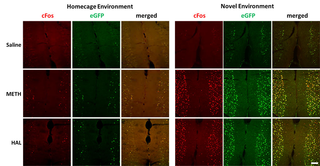
Figure 2. Activation of endogenous cFos expression and eGFP expression in prefrontal cortex. Male LE-Tg(cFos-eGFP)2Ottc rats were injected with saline, methamphetamine (METH; 2.5 mg/kg) or haloperidol (HAL; 1 mg/kg) and immediately placed into home cage or novel environment. Two hours later, animals were perfused with 4% PFA and cryosectioned (40 μm). Sections were immunostained for cFos overnight at 4C. Two hours of secondary antibody. Immunolabeled sections were mounted on slides and imaged for eGFP fluorescence and cFos immunoreactivity using NIKON upright microscope equipped with epifluorescence. Scale bar – 100 μm. Download print resolution version here.

Figure 3. Activation of endogenous cFos expression and eGFP expression in dorsal striatum.

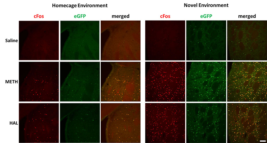
Figure 3.  Activation of endogenous cFos expression and eGFP expression in dorsal striatum.  Male LE-Tg(cFos-eGFP)2Ottc rats were injected with saline, methamphetamine (METH; 2.5 mg/kg) or haloperidol (HAL; 1 mg/kg) and immediately placed into home cage or novel environment. Two hours later, animals were perfused with 4% PFA and cryosectioned (40 μm).  Sections were immunostained for cFos overnight at 4C.  Two hours of secondary antibody.  Immunolabeled sections were mounted on slides and imaged for eGFP fluorescence and cFos immunoreactivity using NIKON upright microscope equipped with epifluorescence.  Scale bar – 100 μm. Download print resolution version here.

Figure 4. Activation of endogenous cFos expression and eGFP expression in hippocampus.

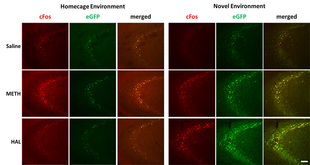
Figure 4.  Activation of endogenous cFos expression and eGFP expression in hippocampus.  Male LE-Tg(cFos-eGFP)2Ottc rats were injected with saline, methamphetamine (METH; 2.5 mg/kg) or haloperidol (HAL; 1 mg/kg) and immediately placed into home cage or novel environment. Two hours later, animals were perfused with 4% PFA and cryosectioned (40 μm).  Sections were immunostained for cFos overnight at 4C.  Two hours of secondary antibody.  Immunolabeled sections were mounted on slides and imaged for eGFP fluorescence and cFos immunoreactivity using NIKON upright microscope equipped with epifluorescence.  Scale bar – 100 μm. Download print resolution version here.

Experiments conducted at the NIDA IRP by Bruce Hope, YaJun Zhang and Brandon Harvey.

Additional phenotypic characterization is recommended for brain region and experimental paradigm of interest.

Breeding Strategy

Breeding Information, click here for PDF

Genotyping Assays

Protocol for genotyping transgenic rats, click here for PDF

References that cite this rat

2019

Demaestri, Camila; Brenhouse, Heather C; Honeycutt, Jennifer A

22 kHz and 55 kHz ultrasonic vocalizations differentially influence neural and behavioral outcomes: Implications for modeling anxiety via auditory stimuli in the rat Journal Article

In: Behavioural Brain Research, vol. 360, pp. 134 - 145, 2019, ISSN: 0166-4328.

Abstract | Links

@article{DEMAESTRI2019134,
title = {22 kHz and 55 kHz ultrasonic vocalizations differentially influence neural and behavioral outcomes: Implications for modeling anxiety via auditory stimuli in the rat},
author = {Camila Demaestri and Heather C Brenhouse and Jennifer A Honeycutt},
url = {https://pubmed.ncbi.nlm.nih.gov/30521931/},
doi = {https://doi.org/10.1016/j.bbr.2018.12.005},
issn = {0166-4328},
year = {2019},
date = {2019-01-01},
journal = {Behavioural Brain Research},
volume = {360},
pages = {134 - 145},
abstract = {The communicative role of ultrasonic vocalizations (USVs) in rats is well established, with distinct USVs indicative of different affective states. USVs in the 22 kHz range are typically emitted by adult rats when in anxiety- or fear-provoking situations (e.g. predator odor, social defeat), while 55 kHz range USVs are typically emitted in appetitive situations (e.g., play, anticipation of reward). Previous work indicates that USVs (real-time and playback) can effectively communicate these affective states and influence changes in behavior and neural activity of the receiver. Changes in cFos activation following 22 kHz USVs have been seen in cortical and limbic regions involved in anxiety, including the basolateral amygdala (BLA). However, it is unclear how USV playback influences cFos activity within the bed nucleus of the stria terminalis (BNST), a region also thought to be critical in processing anxiety-related information, and the nucleus accumbens, a region associated with reward. The present work sought to characterize distinct behavioral, physiological, and neural responses in rats presented with aversive (22 kHz) compared to appetitive (55 kHz) USVs or silence. Our findings show that rats exposed to 22 kHz USVs: 1) engage in anxiety-like behaviors in the elevated zero maze, and 2) show distinct patterns of cFos activation within the BLA and BNST that contrast those seen in 55 kHz playback and silence. Specifically, 22 kHz USVs increased cFos density in the anterodorsal nuclei, while 55 kHz playback increased cFos in the oval nucleus of the BNST, without significant changes within the nucleus accumbens. These results provide important groundwork for leveraging ethologically-relevant stimuli in the rat to improve our understanding of anxiety-related responses in both typical and pathological populations.},
keywords = {},
pubstate = {published},
tppubtype = {article}
}

Close

The communicative role of ultrasonic vocalizations (USVs) in rats is well established, with distinct USVs indicative of different affective states. USVs in the 22 kHz range are typically emitted by adult rats when in anxiety- or fear-provoking situations (e.g. predator odor, social defeat), while 55 kHz range USVs are typically emitted in appetitive situations (e.g., play, anticipation of reward). Previous work indicates that USVs (real-time and playback) can effectively communicate these affective states and influence changes in behavior and neural activity of the receiver. Changes in cFos activation following 22 kHz USVs have been seen in cortical and limbic regions involved in anxiety, including the basolateral amygdala (BLA). However, it is unclear how USV playback influences cFos activity within the bed nucleus of the stria terminalis (BNST), a region also thought to be critical in processing anxiety-related information, and the nucleus accumbens, a region associated with reward. The present work sought to characterize distinct behavioral, physiological, and neural responses in rats presented with aversive (22 kHz) compared to appetitive (55 kHz) USVs or silence. Our findings show that rats exposed to 22 kHz USVs: 1) engage in anxiety-like behaviors in the elevated zero maze, and 2) show distinct patterns of cFos activation within the BLA and BNST that contrast those seen in 55 kHz playback and silence. Specifically, 22 kHz USVs increased cFos density in the anterodorsal nuclei, while 55 kHz playback increased cFos in the oval nucleus of the BNST, without significant changes within the nucleus accumbens. These results provide important groundwork for leveraging ethologically-relevant stimuli in the rat to improve our understanding of anxiety-related responses in both typical and pathological populations.

Close

  • https://pubmed.ncbi.nlm.nih.gov/30521931/
  • doi:https://doi.org/10.1016/j.bbr.2018.12.005

Close

Blog/Comments/Reviews

Last Updated on November 12, 2024

LE-Tg(cFos-eGFP)2Ottc
There are 4 surveyed reports for the receiving and usage of the Long Evans Transgenic cFos-eGFP rats reported an issue during the breeding process.

General Health
There are no reports of general health issues with the LE-Tg(cFos-eGFP)2Ottc rats

Weight
There are no reports of general health issues with the LE-Tg(cFos-eGFP)2Ottc rats

Breeding
One laboratory noticed that the breeding took longer than usual, and they did not receive their first litter until 2-3 months after breeding the LE transgenic male and LE wildtype female rat. This issue only occurred in the F0 within their breeding colony, and it is speculated that the long-distance transport of the transgenics (> 5000 km) may have influenced the breeding delay.

Expression
There are no reports of expression issues with the LE-Tg(cFos-eGFP)2Ottc rats

Other related rats

 LE-Tg(cFos-eGFP)1Ottc  (aka “c-Fos-GFP” rat)
Published as the “c-Fos-GFP” rat which is also a randomly integrated transgene on a Long Evans background that is described by Cifani et al 2012 J Neurosci.  If you are interested in the “c-Fos-GFP” rat, please contact Bruce Hope (bhope@intra.nida.nih.gov).

Acknowledgements

YaJun Zhang, Julie Necarsulmer, Christopher T. Richie, Brandon Harvey, Janette Lebron


Primary Sidebar

Transgenic Rats Project

  • Transgenic Rat Home
  • OTTC Information
  • Publications
  • Technology Development Initiative
  • Links

Organization

  • Organization
  • Faculty
  • Office of the Scientific Director
  • Office of the Clinical Director
  • Administrative Management Branch
  • Molecular Targets and Medications Discovery Branch
  • Cellular and Neurocomputational Systems Branch
  • Molecular Neuropsychiatry Research Branch
  • Neuroimaging Research Branch
  • Behavioral Neuroscience Research Branch
  • Integrative Neuroscience Research Branch
  • Translational Addiction Medicine Branch
  • Core Facilities
  • Careers at NIDA IRP
  • Technology Development Initiative
  • Community Outreach Group
Home / Organization / Office of the Scientific Director / Transgenic Rat Project / LE-Tg(cFos-eGFP)2Ottc
  • National Institute on Drug Abuse
  • NIH Intramural Research Program
  • National Institutes of Health
  • Health and Human Services
  • USA.GOV
  • Emergency Contacts
  • Employee Assistance
  • Treatment Information
  • Contact Us
  • Careers at NIDA IRP
  • Accessibility
  • Privacy
  • HHS Vulnerability Disclosure
  • Freedom of Information Act
  • Document Viewing Tools
  • Offsite Links

  • Home
  • News
    ▼
    • Featured Paper of the Month
    • Reviews to Read
    • Hot off the Press
    • IRP News
    • Awards
    • Technology Development Initiative Paper of the Month
    • Seminar Series
    • Addiction Grand Rounds
  • About
    ▼
    • About NIDA IRP
    • Contact Us
    • Directions and Map
    • Careers at NIDA IRP
    • Emergency Contacts
    • Employee Assistance Resources
  • Organization
    ▼
    • Faculty
    • Office of the Scientific Director
    • Office of the Clinical Director
    • Office of Education and Career Development
    • Administrative Management Branch
    • Molecular Targets and Medications Discovery Branch
    • Cellular and Neurocomputational Systems Branch
    • Molecular Neuropsychiatry Research Branch
    • Neuroimaging Research Branch
    • Behavioral Neuroscience Research Branch
    • Integrative Neuroscience Research Branch
    • Translational Addiction Medicine Branch
    • Core Facilities
    • Community Outreach Group
  • Training Programs
    ▼
    • Office of Education and Career Development
    • OECD Awards
    • Summer Internship Program
    • Postbaccalaureate Program
    • Graduate Partnership Program
    • Postdoctoral Program
    • NIDA Speakers Bureau
    • Clinical Electives Program
    • Clinical Mentoring Program
  • Study Volunteers