Hot Off the Press – June 15, 2022
 Published in Nature Medicine, with contributions by Khaled Moussawi, M.D., Ph.D., Harshwardhan Deshpande, Ph.D., Thomas Ross, Ph.D. and Elliot Stein, Ph.D. of NIDA-IRP.
Summary
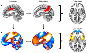
Sometimes regional brain damage (also called a lesion – for example from a stroke) leads to spontaneous remission of addiction – in the present study cigarette smoking. However, if you look at where these lesions occur (compared to lesions that do not lead to remission), there is little overlap. It is possible, then, that the remission was not directly attributable to the location of the lesion, but rather to one or more brain regions connected with (and thus affected by) the lesion. To investigate this, a team of scientists, including three from the NIDA IRP, used a functional MRI (fMRI) technique called ‘functional connectivity.’ Whole-brain functional connectivity maps were generated using the lesion locations as seeds and these maps were compared between remission-causing lesions and non-remission-causing lesions. Given that we lack fMRI data in the patients with lesions, and that the lesions would cause connectivity alterations, the resting state analyses were performed in ‘normal’ samples – both using data from a public dataset and, additionally, a cohort of ‘healthy smokers.’ In contrast to the lack of lesion overlap, there were significant differences between the connectivity maps generated from the 2 groups, and these differences were independent of which ‘normal’ cohort that was used to generate the maps. These differences were centered around brain areas thought to be involved in addiction: bilateral insula, anterior cingulate cortex and medial prefrontal cortex. Finally, aspects of the analyses generalized to data from an alcohol-using population, suggesting a level of common neuronal processing across different drug-induced addictions.
Publication Information
Brain lesions disrupting addiction map to a common human brain circuit Journal Article
In: Nat Med, 2022, ISSN: 1546-170X.
