• Skip to primary navigation
  • Skip to main content
  • Skip to primary sidebar

NIDA IRP

National Institute on Drug Abuse - Intramural Research Program

  National Institute on Drug Abuse | NIH IRP | Treatment Info | Emergency Contacts
  • Home
  • News
    • Featured Paper of the Month
    • Reviews to Read
    • Hot off the Press
    • IRP News
    • Awards
    • Technology Development Initiative Paper of the Month
    • Seminar Series
    • Addiction Grand Rounds
  • About
    • About NIDA IRP
    • Contact Us
    • Directions and Map
    • Careers at NIDA IRP
    • Emergency Contacts
    • Employee Assistance Resources
  • Organization
    • Faculty
    • Office of the Scientific Director
    • Office of the Clinical Director
    • Office of Education and Career Development
    • Administrative Management Branch
    • Molecular Targets and Medications Discovery Branch
    • Cellular and Neurocomputational Systems Branch
    • Molecular Neuropsychiatry Research Branch
    • Neuroimaging Research Branch
    • Behavioral Neuroscience Research Branch
    • Integrative Neuroscience Research Branch
    • Translational Addiction Medicine Branch
    • Core Facilities
    • Community Outreach Group
  • Training Programs
    • Office of Education and Career Development
    • OECD Awards
    • Summer Internship Program
    • Postbaccalaureate Program
    • Graduate Partnership Program
    • Postdoctoral Program
    • NIDA Speakers Bureau
    • Clinical Electives Program
    • Clinical Mentoring Program
  • Study Volunteers

Flow Cytometry of Synaptoneurosomes (FCS) Reveals Increased Ribosomal S6 and Calcineurin Proteins in Activated Medial Prefrontal Cortex to Nucleus Accumbens Synapses

Study author Javier Rubio (left) and a figure from this article (right)Hot Off the Press – June 27, 2023

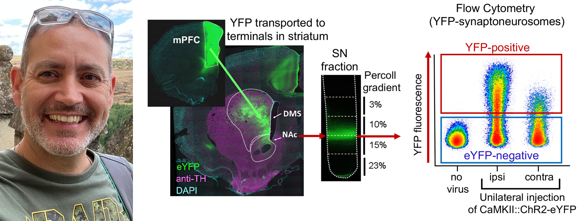
Published in The Journal of Neuroscience by Javier Rubio and Bruce Hope, et al. of the NIDA IRP Neuronal Ensembles in Drug Addiction Section.

The study is a novel approach to study molecular alterations in individual synapses using Flow Cytometry of Synaptoneurosomes (FCS). The FCS approach was used to identify activity-dependent increases of ribosomal protein S6 in post-synaptic endings and calcineurin in presynaptic terminals coming from mPFC to nucleus accumbens in rats. S6 and calcineurin may be useful markers to identify activated synaptic ensembles similar to how Fos is used as a marker to identify activated cell bodies in neuronal ensembles to study learning-induced plasticity changes. This project was a collaboration with several groups from NIDA-IRP and NIA-IRP, especially the participation of the Flow Cytometry Unit lead by Chris Dunn at NIA and the Confocal and Electron Microscopy Core lead by Steven Zhang at NIDA.

Publication Information

Rubio, F Javier; Olivares, Daniel E; Dunn, Christopher; Zhang, Shiliang; Hilaire, Elias M; Henry, Akeem; Mejias-Aponte, Carlos; Nogueras-Ortiz, Carlos J; Selvam, Pooja V; Cruz, Fabio C; Madangopal, Rajtarun; Morales, Marisela; Hope, Bruce T

Flow Cytometry of Synaptoneurosomes (FCS) Reveals Increased Ribosomal S6 and Calcineurin Proteins in Activated Medial Prefrontal Cortex to Nucleus Accumbens Synapses Journal Article

In: J Neuroci., vol. 43, iss. 23, pp. 4217-4233, 2023.

Abstract | Links

@article{12023,
title = {Flow Cytometry of Synaptoneurosomes (FCS) Reveals Increased Ribosomal S6 and Calcineurin Proteins in Activated Medial Prefrontal Cortex to Nucleus Accumbens Synapses},
author = {F Javier Rubio and Daniel E Olivares and Christopher Dunn and Shiliang Zhang and Elias M Hilaire and Akeem Henry and Carlos Mejias-Aponte and Carlos J Nogueras-Ortiz and Pooja V Selvam and Fabio C Cruz and Rajtarun Madangopal and Marisela Morales and Bruce T Hope},
url = {https://pubmed.ncbi.nlm.nih.gov/37160369/},
doi = {10.1523/JNEUROSCI.0927-22.2023},
year = {2023},
date = {2023-06-07},
urldate = {2023-06-07},
journal = {J Neuroci.},
volume = {43},
issue = {23},
pages = {4217-4233},
abstract = {Learning and behavior activate cue-specific patterns of sparsely distributed cells and synapses called ensembles that undergo memory-encoding engram alterations. While Fos is often used to label selectively activated cell bodies and identify neuronal ensembles, there is no comparable endogenous marker to label activated synapses and identify synaptic ensembles. For the purpose of identifying candidate synaptic activity markers, we optimized a flow cytometry of synaptoneurosome (FCS) procedure for assessing protein alterations in activated synapses from male and female rats. After injecting yellow fluorescent protein (YFP)-expressing adeno-associated virus into medial prefrontal cortex (mPFC) to label terminals in nucleus accumbens (NAc) of rats, we injected 20 mg/kg cocaine in a novel context (cocaine+novelty) to activate synapses, and prepared NAc synaptoneurosomes 0-60 min following injections. For FCS, we used commercially available antibodies to label presynaptic and postsynaptic markers synaptophysin and PSD-95 as well as candidate markers of synaptic activity [activity-regulated cytoskeleton protein (Arc), CaMKII and phospho-CaMKII, ribosomal protein S6 (S6) and phospho-S6, and calcineurin and phospho-calcineurin] in YFP-labeled synaptoneurosomes. Cocaine+novelty increased the percentage of S6-positive synaptoneurosomes at 5-60 min and calcineurin-positive synaptoneurosomes at 5-10 min. Electron microscopy verified that S6 and calcineurin levels in synaptoneurosomes were increased 10 min after cocaine+novelty. Pretreatment with the anesthetic chloral hydrate blocked cocaine+novelty-induced S6 and calcineurin increases in synaptoneurosomes, and novel context exposure alone (without cocaine) increased S6, both of which indicate that these increases were due to neural activity per se. Overall, FCS can be used to study protein alterations in activated synapses coming from specifically labeled mPFC projections to NAc.SIGNIFICANCE STATEMENT Memories are formed during learning and are stored in the brain by long-lasting molecular and cellular alterations called engrams formed within specific patterns of cue-activated neurons called neuronal ensembles. While Fos has been used to identify activated ensemble neurons and the engrams within them, we have not had a similar marker for activated synapses that can be used to identify synaptic engrams. Here we developed a procedure for high-throughput in-line analysis of flow cytometry of synaptoneurosome (FCS) and found that ribosomal S6 protein and calcineurin were increased in activated mPFC-NAc synapses. FCS can be used to study protein alterati},
keywords = {},
pubstate = {published},
tppubtype = {article}
}

Close

Learning and behavior activate cue-specific patterns of sparsely distributed cells and synapses called ensembles that undergo memory-encoding engram alterations. While Fos is often used to label selectively activated cell bodies and identify neuronal ensembles, there is no comparable endogenous marker to label activated synapses and identify synaptic ensembles. For the purpose of identifying candidate synaptic activity markers, we optimized a flow cytometry of synaptoneurosome (FCS) procedure for assessing protein alterations in activated synapses from male and female rats. After injecting yellow fluorescent protein (YFP)-expressing adeno-associated virus into medial prefrontal cortex (mPFC) to label terminals in nucleus accumbens (NAc) of rats, we injected 20 mg/kg cocaine in a novel context (cocaine+novelty) to activate synapses, and prepared NAc synaptoneurosomes 0-60 min following injections. For FCS, we used commercially available antibodies to label presynaptic and postsynaptic markers synaptophysin and PSD-95 as well as candidate markers of synaptic activity [activity-regulated cytoskeleton protein (Arc), CaMKII and phospho-CaMKII, ribosomal protein S6 (S6) and phospho-S6, and calcineurin and phospho-calcineurin] in YFP-labeled synaptoneurosomes. Cocaine+novelty increased the percentage of S6-positive synaptoneurosomes at 5-60 min and calcineurin-positive synaptoneurosomes at 5-10 min. Electron microscopy verified that S6 and calcineurin levels in synaptoneurosomes were increased 10 min after cocaine+novelty. Pretreatment with the anesthetic chloral hydrate blocked cocaine+novelty-induced S6 and calcineurin increases in synaptoneurosomes, and novel context exposure alone (without cocaine) increased S6, both of which indicate that these increases were due to neural activity per se. Overall, FCS can be used to study protein alterations in activated synapses coming from specifically labeled mPFC projections to NAc.SIGNIFICANCE STATEMENT Memories are formed during learning and are stored in the brain by long-lasting molecular and cellular alterations called engrams formed within specific patterns of cue-activated neurons called neuronal ensembles. While Fos has been used to identify activated ensemble neurons and the engrams within them, we have not had a similar marker for activated synapses that can be used to identify synaptic engrams. Here we developed a procedure for high-throughput in-line analysis of flow cytometry of synaptoneurosome (FCS) and found that ribosomal S6 protein and calcineurin were increased in activated mPFC-NAc synapses. FCS can be used to study protein alterati

Close

  • https://pubmed.ncbi.nlm.nih.gov/37160369/
  • doi:10.1523/JNEUROSCI.0927-22.2023

Close

Primary Sidebar

News

  • All News and Featured Publications
  • Featured Paper of the Month
  • Hot off the Press
  • Reviews to Read
  • IRP News
  • Awards
  • Technology Development Initiative Paper of the Month
  • Seminar Series
Home / News Main / Hot off the Press / Flow Cytometry of Synaptoneurosomes (FCS) Reveals Increased Ribosomal S6 and Calcineurin Proteins in Activated Medial Prefrontal Cortex to Nucleus Accumbens Synapses
  • National Institute on Drug Abuse
  • NIH Intramural Research Program
  • National Institutes of Health
  • Health and Human Services
  • USA.GOV
  • Emergency Contacts
  • Employee Assistance
  • Treatment Information
  • Contact Us
  • Careers at NIDA IRP
  • Accessibility
  • Privacy
  • HHS Vulnerability Disclosure
  • Freedom of Information Act
  • Document Viewing Tools
  • Offsite Links

  • Home
  • News
    ▼
    • Featured Paper of the Month
    • Reviews to Read
    • Hot off the Press
    • IRP News
    • Awards
    • Technology Development Initiative Paper of the Month
    • Seminar Series
    • Addiction Grand Rounds
  • About
    ▼
    • About NIDA IRP
    • Contact Us
    • Directions and Map
    • Careers at NIDA IRP
    • Emergency Contacts
    • Employee Assistance Resources
  • Organization
    ▼
    • Faculty
    • Office of the Scientific Director
    • Office of the Clinical Director
    • Office of Education and Career Development
    • Administrative Management Branch
    • Molecular Targets and Medications Discovery Branch
    • Cellular and Neurocomputational Systems Branch
    • Molecular Neuropsychiatry Research Branch
    • Neuroimaging Research Branch
    • Behavioral Neuroscience Research Branch
    • Integrative Neuroscience Research Branch
    • Translational Addiction Medicine Branch
    • Core Facilities
    • Community Outreach Group
  • Training Programs
    ▼
    • Office of Education and Career Development
    • OECD Awards
    • Summer Internship Program
    • Postbaccalaureate Program
    • Graduate Partnership Program
    • Postdoctoral Program
    • NIDA Speakers Bureau
    • Clinical Electives Program
    • Clinical Mentoring Program
  • Study Volunteers